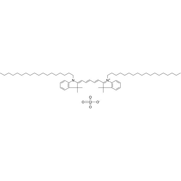
DiD perchlorate; 纯度: 98.96%
DiD perchlorate 是一种远红色荧光亲脂性花青染料。DiD perchlorate 能快速稳定地与磷脂细胞膜结合,能用于细胞追踪。

DiD perchlorate Chemical Structure
CAS No. : 127274-91-3
| 规格 | 价格 | 是否有货 | 数量 |
|---|---|---|---|
| 5 mg | ¥1200 | In-stock | |
| 10 mg | ; | 询价 | ; |
| 50 mg | ; | 询价 | ; |
* Please select Quantity before adding items.
DiD perchlorate 相关产品
bull;相关化合物库:
- Bioactive Compound Library Plus
| 生物活性 |
DiD perchlorate is a far-red fluorescent lipophilic cyanine dye. DiD perchlorate can rapidly and stably integrate into the phospholipid cell membrane. DiD perchlorate is used to cells tracking[1][2][3]. |
||||||||||||||||
|---|---|---|---|---|---|---|---|---|---|---|---|---|---|---|---|---|---|
| 体内研究 (In Vivo) |
Two weeks after injection of stained cells, a single, bright, DiD perchlorate (DiD)-positive cells located between 15 to 40 µm from the endosteum is found. Results reveal a progressive appearance of cell clusters of decreased dye intensity, consistent with the partitioning of DiD perchlorate label on cell division[3]. MCE has not independently confirmed the accuracy of these methods. They are for reference only. |
||||||||||||||||
| 分子量 |
959.90 |
||||||||||||||||
| Formula |
C61H99ClN2O4 |
||||||||||||||||
| CAS 号 |
127274-91-3 |
||||||||||||||||
| 运输条件 |
Room temperature in continental US; may vary elsewhere. |
||||||||||||||||
| 储存方式 |
4deg;C, protect from light, stored under nitrogen *该产品在溶液状态不稳定,建议您现用现配,即刻使用。 |
||||||||||||||||
| 溶解性数据 |
In Vitro:;
DMSO : 25 mg/mL (26.04 mM; ultrasonic and warming and heat to 60°C) 配制储备液
*
请根据产品在不同溶剂中的溶解度,选择合适的溶剂配制储备液;该产品在溶液状态不稳定,建议您现用现配,即刻使用。 In Vivo:
请根据您的实验动物和给药方式选择适当的溶解方案。以下溶解方案都请先按照 In Vitro 方式配制澄清的储备液,再依次添加助溶剂: ——为保证实验结果的可靠性,澄清的储备液可以根据储存条件,适当保存;体内实验的工作液,建议您现用现配,当天使用; 以下溶剂前显示的百
|
||||||||||||||||
| 参考文献 |
|
| Animal Administration [1] |
1 to 5×10⁵ cells per mL HSPCs are stained with 5 μM DiD perchlorate (DiD) or 7.5 μM DiI in PBS without serum for 10 min at 37°C, washed once in PBS and immediately injected into the tail vein of recipient mice. Unless stated differently, each imaged CD45.2 mouse receive 8,000 to 15,000 labelled CD45.1 HSPCs together with 3 to 5×10⁵ CD45.2 supportive whole bone-marrow mononuclear cells to ensure survival[1]. MCE has not independently confirmed the accuracy of these methods. They are for reference only. |
|---|---|
| 参考文献 |
|
