yamato雅马拓台式超音波清洗机

yamato雅马拓台式超音波清洗机

yamato雅马拓台式超音波清洗机

产品编号:
市场价:¥0.00
会员价:¥0.00
品牌:YAMATO 雅马拓
生产厂家:yamato雅马拓
需求数量:

  • 产品概览
  • 技术参数
  • 订购信息
  • 相关资料
  • 相关产品
控制器部分 MT机型
●定时:60分钟设定可能(右回旋)
●保持:连续运行(左回旋)
●MTH机型:附有加热器开关
DTH机型
1: POWER 开关
2: ON/OFF 开关
3: LED 显示
4: SET 显示
5: CLEAR 显示
6: SELECT OPTION 键
7: FUNCTION INDICATOR


  • 频率带狭窄,对固着物的清洗能力超群。另外对固体 样品的溶解、精制等也有很好的效果。
  • MTH/DTH型附有加热器,可以让清洗剂和清洗液快速的 混合,超音波清洗效果更好。 专用清洗剂有用来清洗锈迹、金属附着物最为合适的酸性 洗剂和碱性洗剂。 加装水槽盖可以更快的使清洗液温度上升。
  • 新的清洗液中有溶解进去的气体(主要是空气),会 妨碍清洗液的气穴。通过除去瓦斯功能在清洗前加以去 处的话,就能更好的提高超音波效果。 DTH机型标准装备Degas运行模式。


  • 品号 型号 功能 外形尺寸(mm)
    (WxDxH)
    内槽尺寸(mm)
    (WxDxH)
    容量 (L) 重量 (kg) 频率 (kHz) 功率 (W) 加热 (W) 定时 (min)
    291280 1510J-MT 机械定时 254*305*295 150*135*102 2.0 3.5 42 90 0~60
    291281 1510J-MTH 机械定时
    /加热
      65 0~60
    291267 1510J-DTH 数码控制/
    温调加热/
    定时/除瓦斯
      65 0~99
    291282 2510J-MT 机械定时 338*305*295 235*135*102 2.8 4.2 42 125 0~60
    291283 2510J-MTH 机械定时
    /加热
      115 0~60
    291270 2510J-DTH 数码控制/
    温调加热/
    定时/除瓦斯
      115 0~99
    291284 3510J-MT 机械定时 399*318*371 292*152*152 5.7 6.5 42 130 0~60
    291285 3510J-MTH 机械定时
    /加热
      200 0~60
    291273 3510J-DTH 数码控制/
    温调加热/
    定时/除瓦斯
      200 0~99
    291286 5510J-MT 机械定时 399*401*371 292*241*152 9.5 7.6 42 180 0~60
    291287 5510J-MTH 机械定时
    /加热
      290 0~60
    291276 5510J-DTH 数码控制/
    温调加热/
    定时/除瓦斯
      290 0~99
    291288 8510J-MT 机械定时 599*467*376 495*292*152 20.1 12.0 44 320 0~60
    291289 8510J-MTH 机械定时
    /加热
      560 0~60
    291279 8510J-DTH 数码控制/
    温调加热/
    定时/除瓦斯
      560 0~99

    荧光染料SBFI-AM

    荧光染料Dye Reagents SBFI-AM;

    SBFI-AM 是一种 Na+ 选择性的荧光指示剂。SBFI-AM 显示出对 Na+ 的选择性高于对 K+ 的选择性。

    SBFI-AM

    SBFI-AM Chemical Structure

    CAS No. : 129423-53-6

    规格 是否有货
    100 mg ; 询价 ;
    250 mg ; 询价 ;
    500 mg ; 询价 ;

    * Please select Quantity before adding items.

    生物活性

    SBFI-AM is a Na+ selective fluorescent indicator. SBFI-AM shows selectivity for Na+ over K+[1].

    分子量

    1127.06

    Formula

    C56H58N2O23

    CAS 号

    129423-53-6

    运输条件

    Room temperature in continental US; may vary elsewhere.

    储存方式

    Please store the product under the recommended conditions in the Certificate of Analysis.

    参考文献
    • [1]. Minta A, et, al. Fluorescent indicators for cytosolic sodium. J Biol Chem. 1989 Nov 15;264(32):19449-57.

    BAPTA-AM

    生化分析试剂 Biochemical Assay Reagents
    BAPTA-AM; 纯度: 99.23%

    BAPTA-AM 是一种可透过细胞膜的钙螯合剂 (Ca2+ chelator)。在 HEK 293 细胞中,BAPTA-AM 阻断 hERGhKv1.3hKv1.5 通道,IC50 分别为 1.3,1.45 和 1.23 μM。

    BAPTA-AM

    BAPTA-AM Chemical Structure

    CAS No. : 126150-97-8

    规格 价格 是否有货 数量
    10;mM;*;1 mL in DMSO ¥1260 In-stock
    5 mg ¥900 In-stock
    10 mg ¥1500 In-stock
    50 mg ¥4500 In-stock
    100 mg ; 询价 ;
    200 mg ; 询价 ;

    * Please select Quantity before adding items.

    BAPTA-AM 相关产品

    bull;相关化合物库:

    • Bioactive Compound Library Plus
    • Membrane Transporter/Ion Channel Compound Library
    • Anti-Cardiovascular Disease Compound Library
    • Anti-Alzheimer’s Disease Compound Library
    • Neurodegenerative Disease-related Compound Library

    生物活性

    BAPTA-AM is a well-known membrane permeable Ca2+ chelator. BAPTA-AM inhibits hERG channels, hKv1.3 and hKv1.5 channels in HEK 293 cells with IC50s of 1.3 μM, 1.45 μM and 1.23 μM, respectively[1].

    IC50 Target

    Ca2+ chelator[1]
    IC50: 1.3 μM (hERG channel, in HEK 293 cells), 1.45 μM (hKv1.3, in HEK 293 cells), 1.23 μM (hKv1.5, in HEK 293 cells)[1]

    体外研究
    (In Vitro)

    BAPTA-AM inhibits neuronal Ca2+-activated K+ channel currents, and up-regulates the decreased cardiac sodium current (INa) density by chelating intracellular Ca2+[1].
    BAPTA-AM (BAPTA/AM), an intracellular calcium chelator, induces delayed necrosis by lipoxygenase-mediated free radicals in mouse cortical cultures. BAPTA-AM prevents free radical-mediated toxicity promote apoptosis in non-neuronal cells and produce a beneficial effect in neuronal cells by protecting neurons from ischemic damage. In addition, it has been suggested that BAPTA-AM induces a late, but not early, increase of intracellular calcium in I-IL-60 neoplastic cells. Mixed cortical cell cultures (DIV 13-16) exposed to 10 μM BAPTA-AM for 24- or 48-hr show moderate (45-70%) neuronal injury as evaluated by increased LDH release into the bathing medium after 24-48-hr. Exposure of cortical cultures to 3-10 μM BAPTA-AM for 48-hr evoke dose-dependent neuronal damage[2].

    MCE has not independently confirmed the accuracy of these methods. They are for reference only.

    分子量

    764.68

    Formula

    C34H40N2O18

    CAS 号

    126150-97-8

    运输条件

    Room temperature in continental US; may vary elsewhere.

    储存方式
    Powder -20deg;C 3 years
    4deg;C 2 years
    In solvent -80deg;C 6 months
    -20deg;C 1 month
    溶解性数据
    In Vitro:;

    DMSO : 50 mg/mL (65.39 mM; Need ultrasonic)

    H2O : < 0.1 mg/mL (ultrasonic) (insoluble)

    配制储备液
    浓度 溶剂体积 质量 1 mg 5 mg 10 mg
    1 mM 1.3077 mL 6.5387 mL 13.0774 mL
    5 mM 0.2615 mL 1.3077 mL 2.6155 mL
    10 mM 0.1308 mL 0.6539 mL 1.3077 mL

    *

    请根据产品在不同溶剂中的溶解度选择合适的溶剂配制储备液;一旦配成溶液,请分装保存,避免反复冻融造成的产品失效
    储备液的保存方式和期限:-80°C, 6 months; -20°C, 1 month。-80°C 储存时,请在 6 个月内使用,-20°C 储存时,请在 1 个月内使用。

    In Vivo:

    请根据您的实验动物和给药方式选择适当的溶解方案。以下溶解方案都请先按照 In Vitro 方式配制澄清的储备液,再依次添加助溶剂:

    ——为保证实验结果的可靠性,澄清的储备液可以根据储存条件,适当保存;体内实验的工作液,建议您现用现配,当天使用; 以下溶剂前显示的百
    分比是指该溶剂在您配制终溶液中的体积占比;如在配制过程中出现沉淀、析出现象,可以通过加热和/或超声的方式助溶

    • 1.

      请依序添加每种溶剂:;10% DMSO ;; 40% PEG300 ;; 5% Tween-80 ;; 45% saline

      Solubility: ≥ 2.5 mg/mL (3.27 mM); Clear solution

      此方案可获得 ≥ 2.5 mg/mL (3.27 mM,饱和度未知) 的澄清溶液。

      以 1 mL 工作液为例,取 100 μL 25.0 mg/mL 的澄清 DMSO 储备液加到 400 μL PEG300 中,混合均匀;向上述体系中加入50 μL Tween-80,混合均匀;然后继续加入 450 μL生理盐水定容至 1 mL。

      将 0.9 g 氯化钠,完全溶解于 100 mL ddH₂O 中,得到澄清透明的生理盐水溶液

    • 2.

      请依序添加每种溶剂:;10% DMSO ;; 90% (20% SBE-β-CD in saline)

      Solubility: 2.5 mg/mL (3.27 mM); Suspended solution; Need ultrasonic

      此方案可获得 2.5 mg/mL (3.27 mM) 的均匀悬浊液,悬浊液可用于口服和腹腔注射。

      以 1 mL 工作液为例,取 100 μL 25.0 mg/mL 的澄清 DMSO 储备液加到 900 μL 20% 的 SBE-β-CD 生理盐水水溶液中,混合均匀。

      将 2 g 磺丁基醚 β-环糊精加入 5 mL 生理盐水中,再用生理盐水定容至 10 mL,完全溶解,澄清透明
    • 3.

      请依序添加每种溶剂:;10% DMSO ;; 90% corn oil

      Solubility: ≥ 2.5 mg/mL (3.27 mM); Clear solution

      此方案可获得 ≥ 2.5 mg/mL (3.27 mM,饱和度未知) 的澄清溶液,此方案不适用于实验周期在半个月以上的实验。

      以 1 mL 工作液为例,取 100 μL 25.0 mg/mL 的澄清 DMSO 储备液加到 900 μL玉米油中,混合均匀。

    *以上所有助溶剂都可在 MCE 网站选购。
    参考文献
    • [1]. Tang Q, et al. The membrane permeable calcium chelator BAPTA-AM directly blocks human ether a-go-go-related gene potassium channels stably expressed in HEK 293 cells. Biochem Pharmacol. 2007 Dec 3;74(11):1596-607.

      [2]. Wie MB, et al. BAPTA/AM, an intracellular calcium chelator, induces delayed necrosis by lipoxygenase-mediated free radicals in mouse cortical cultures. Prog Neuropsychopharmacol Biol Psychiatry. 2001 Nov;25(8):1641-59.

    Cell Assay
    [1]

    Neuronal injury is quantitatively estimated by measuring lactate dehydrogenase (LDH) released from damaged cells into the bathing medium 24- or 48-hr after the 10 μM BAPTA/AM treatment. The morphological findings are confirmed by staining with neuron-specific enolase (NSE) antibody and tryphan blue[1].

    MCE has not independently confirmed the accuracy of these methods. They are for reference only.

    参考文献
    • [1]. Tang Q, et al. The membrane permeable calcium chelator BAPTA-AM directly blocks human ether a-go-go-related gene potassium channels stably expressed in HEK 293 cells. Biochem Pharmacol. 2007 Dec 3;74(11):1596-607.

      [2]. Wie MB, et al. BAPTA/AM, an intracellular calcium chelator, induces delayed necrosis by lipoxygenase-mediated free radicals in mouse cortical cultures. Prog Neuropsychopharmacol Biol Psychiatry. 2001 Nov;25(8):1641-59.

    Calcein-AM(Synonyms: 钙黄绿素-AM Calcein acetoxymethyl ester)

    Calcein-AM;(Synonyms: 钙黄绿素-AM; Calcein acetoxymethyl ester) 纯度: ge;96.0%

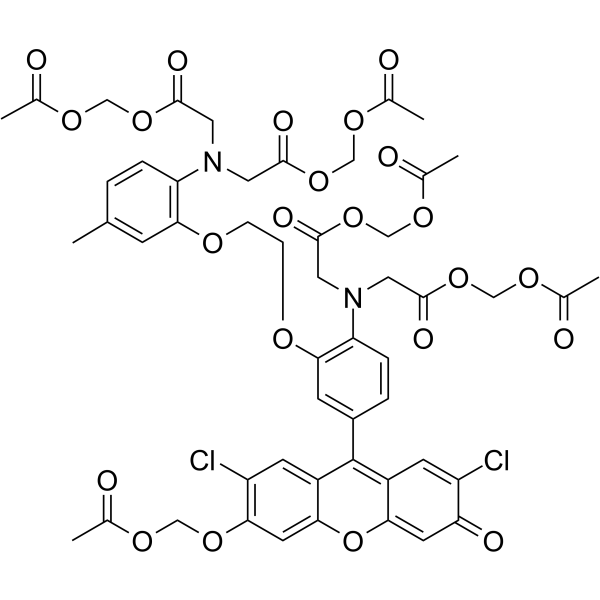
    Calcein-AM是用于测定细胞活力的可以渗透细胞的荧光染料。

    Calcein-AMamp;;(Synonyms: 钙黄绿素-AM; Calcein acetoxymethyl ester)

    Calcein-AM Chemical Structure

    CAS No. : 148504-34-1

    规格 价格 是否有货 数量
    100 μg(2 mg/mL * 50 μL in DMSO) ¥1300 In-stock
    500 μg(2 mg/mL * 250 μL in DMSO) ¥4750 In-stock

    * Please select Quantity before adding items.

    生物活性

    Calcein-AM is cell-permeable fluorescent dye used to determine the cell viability.

    体外研究
    (In Vitro)

    The calcein-AM dye used to stain the living cells is shown to have a low spontaneousleakage rate less than 15% in 4 hours at 37°C. Dilutions of targets stained by calcein-AM has a linear relationship with measured fluorescence values. NK cells, LAKs, and CTLs are readily detectable by this microtest. Quantitation of killing and kinetic analysis is readily performed with the test system[1]. Calcein-AM is pH independent, better retained and more photostable. In addition, the high level of intracellular retention of calcein-AM and its low-level release after incorporation exclude possible cell-monolayer labeling and allow its use in a cell-cell interaction assay. Moreover, the bright fluorescence can easily be detected and measured by a microplate fluorescence reader[2]. Calcein-AM is a highly lipophilic vital dye that rapidly enters viable cells, is converted by intracellular esterases to calcein that produces an intense green (530-nm) signal, and is retained by cells with intact plasma membrane. From dying or damaged cells with compromised membrane integrity or from cells expressing multidrug resistance protein (MRP), unhydrolyzed substrates and their fluorescent products are rapidly extruded from cells. The calcein-AM assay has been used to assess the cell viability, cytotoxicity and tp quantitate apoptosis[3].

    MCE has not independently confirmed the accuracy of these methods. They are for reference only.

    体内研究
    (In Vivo)

    Calcein-AM is found to be suitable for in vivo studies, because it has no deleterious effects on cell function and is, indeed, a marker of cell viability[2].

    MCE has not independently confirmed the accuracy of these methods. They are for reference only.

    分子量

    994.86

    Formula

    C46H46N2O23

    CAS 号

    148504-34-1

    中文名称

    钙黄绿素-AM

    运输条件

    Room temperature in continental US; may vary elsewhere.

    储存方式

    -20deg;C, protect from light

    *In solvent : -80deg;C, 6 months; -20deg;C, 1 month (protect from light)

    参考文献
    • [1]. Wang XM, et al. A new microcellular cytotoxicity test based on calcein-AM release. Hum Immunol. 1993 Aug;37(4):264-70.

      [2]. Braut-Boucher F, et al. A non-isotopic, highly sensitive, fluorimetric, cell-cell adhesion microplate assay using calceinAM-labeled lymphocytes. J Immunol Methods. 1995 Jan 13;178(1):41-51.

      [3]. Bratosin D, et al. Novel fluorescence assay using calcein-AM for the determination of human erythrocyte viabilityand aging. Cytometry A. 2005 Jul;66(1):78-84.

    Cell Assay
    [1][2][3]

    K562, Daudi, and Chang liver cells are labeled with calcein-AM. Calcein-AM’s excitation and emission wavelengths are 496 nm and 520 nm, respectively. The filter/mirror combination used to detect calcein-AM’s green fluorescence includes the 490-nm excitation and 520-nm emission filters with a dichroic mirror. Differences in the automatic fluorescence readings between the test and control wells determine the results[1]. A simple and sensitive cell-cell adhesion microplate assay is established using the calcein-AM. The procedure involves three steps: the labeling of lymphocytes with an adequate concentration of calcein-AM (20 μM) during a short incubation period (30 min); the adhesion of 2×10⁵ labeled lymphocytes per well to confluent keratinocyte or fibroblast monolayers grown in microtiter plates for 90 min; and, finally, measurement of the fluorescent signal utilizing a new system of cold-light microfluorimetry[2]. Cells are incubated for 15 min in 1 mL of a 1% saponin solution in PBS buffer, pH 7.4, containing 0.05% sodium azide. After saponin permeabilization, 4×10⁵ RBCs in suspension in PBS buffer containing 0.1% saponin and 0.05% sodium azide are incubated (37°C in the dark for 45 min) with calcein-AM to a final concentration of 5 μM, ished three times with the same PBS buffer containing 0.1% saponin and 0.05% sodium azide, and the cell viability is analyzed by flow cytometry[3].

    MCE has not independently confirmed the accuracy of these methods. They are for reference only.

    参考文献
    • [1]. Wang XM, et al. A new microcellular cytotoxicity test based on calcein-AM release. Hum Immunol. 1993 Aug;37(4):264-70.

      [2]. Braut-Boucher F, et al. A non-isotopic, highly sensitive, fluorimetric, cell-cell adhesion microplate assay using calceinAM-labeled lymphocytes. J Immunol Methods. 1995 Jan 13;178(1):41-51.

      [3]. Bratosin D, et al. Novel fluorescence assay using calcein-AM for the determination of human erythrocyte viabilityand aging. Cytometry A. 2005 Jul;66(1):78-84.

    Fluo-4 AM

    Fluo-4 AM;

    Fluo-4 AM 是一种常用的检测细胞内 Ca2+ 浓度的探针。

    Fluo-4 AMamp;;

    Fluo-4 AM Chemical Structure

    CAS No. : 273221-67-3

    规格 价格 是否有货
    100 μg ¥1500 询问价格 货期

    * Please select Quantity before adding items.

    生物活性

    Fluo-4 AM is a cell-permeable Ca2+ indicator[1].

    体外研究
    (In Vitro)

    Fluo-4 AM is a fluorescent dye (λex=494 nm, λem=516 nm). Preloaded with Fuo-4 AM, a very bright fluorescence image is observed. In a parallel experiment with fluo-3 AM-loaded cells, the resulting fluorescence image, although clearly discernable in this case, is less bright[1]. Guidelines (Following is our recommended protocol. This protocol only provides a guideline, and should be modified according to your specific needs).
    1. Count the cells and take 106 cells from each sample (control and experiment/s).
    2. Collect the cells (5 min, 3000×g, 4 °C) and wash once in PBS.
    3. Resuspend the cells in 0.5-ml PBS and add 0.5 μl of Fluo-4-AM (1 mM stock) to a final concentration of 1 μM. Incubate at 37 °C for 1 h.
    4. Wash the cells three times (2 min, 3000×g) with PBS and finally resuspend in 1-ml PBS. Separate into 2 flow cytometry tubes—0.5 ml in each.
    5. Evaluate the staining by flow cytometry and analyze the data by a software such as CellQuest software.

    MCE has not independently confirmed the accuracy of these methods. They are for reference only.

    分子量

    1096.94

    Formula

    C51H50F2N2O23

    CAS 号

    273221-67-3

    运输条件

    Room temperature in continental US; may vary elsewhere.

    储存方式

    -20deg;C, sealed storage, away from moisture and light

    *该产品在溶液状态不稳定,建议您现用现配,即刻使用。

    参考文献
    • [1]. Gee KR, et al. Chemical and physiological characterization of fluo-4 Ca(2+)-indicator dyes. Cell Calcium. 2000 Feb;27(2):97-106.

      [2]. Fluo-4.

    Cell Assay
    [1]

    For measuring fluorescence from cells in suspension, dilutions to 2 to 3×10⁶ cells are made, from cultures of rat basophilic leukemia (RBL) cells. Cells are incubated in suspension in 1 μM dye (including Fluo-4 AM) for 30 min at 37°C. Cell suspensions are then transferred to cuvets for measurements of fluorescence emission intensity by spectrofluorometer[1].

    MCE has not independently confirmed the accuracy of these methods. They are for reference only.

    参考文献
    • [1]. Gee KR, et al. Chemical and physiological characterization of fluo-4 Ca(2+)-indicator dyes. Cell Calcium. 2000 Feb;27(2):97-106.

      [2]. Fluo-4.

    荧光染料HKYellow-AM (6/12-mixture)

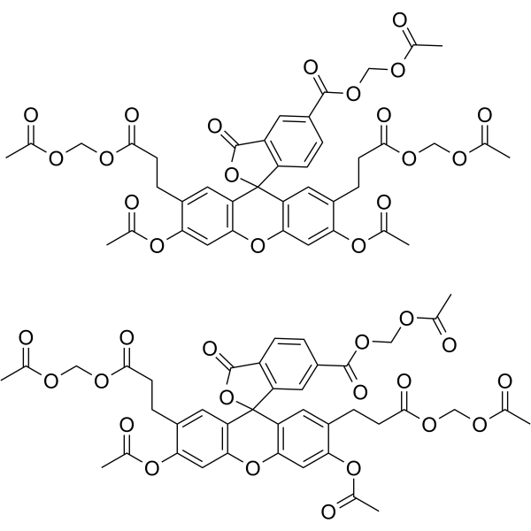
    荧光染料Dye Reagents HKYellow-AM (6/12-mixture); 纯度: 98.69%

    HKYellow-AM (6/12-mixture) 一种荧光探针,可用于过氧亚硝酸盐的灵敏和特异检测。详细信息请参考专利文献 EP2809666B1 中的化合物 14。

    HKYellow-AM (6/12-mixture)amp;;

    HKYellow-AM (6/12-mixture) Chemical Structure

    CAS No. : 1448821-89-3

    规格 价格 是否有货 数量
    1 mg ¥3000 In-stock
    5 mg ¥9000 In-stock
    10 mg ¥15500 In-stock
    50 mg ; 询价 ;
    100 mg ; 询价 ;

    * Please select Quantity before adding items.

    HKYellow-AM (6/12-mixture) 相关产品

    bull;相关化合物库:

    • Bioactive Compound Library Plus

    生物活性

    HKYellow-AM (6/12-mixture) is a fluorogenic probe extracted from patent EP2809666B1, compound 14, which can be used for sensitive and specific detection of peroxynitrite[1].

    分子量

    1422.40

    Formula

    C80H78Cl2N4O16

    CAS 号

    1448821-89-3

    运输条件

    Room temperature in continental US; may vary elsewhere.

    储存方式

    -20deg;C, sealed storage, away from moisture and light

    *In solvent : -80deg;C, 6 months; -20deg;C, 1 month (sealed storage, away from moisture and light)

    溶解性数据
    In Vitro:;

    DMSO : 130 mg/mL (91.39 mM; Need ultrasonic)

    配制储备液
    浓度 溶剂体积 质量 1 mg 5 mg 10 mg
    1 mM 0.7030 mL 3.5152 mL 7.0304 mL
    5 mM 0.1406 mL 0.7030 mL 1.4061 mL
    10 mM 0.0703 mL 0.3515 mL 0.7030 mL

    *

    请根据产品在不同溶剂中的溶解度选择合适的溶剂配制储备液;一旦配成溶液,请分装保存,避免反复冻融造成的产品失效
    储备液的保存方式和期限:-80°C, 6 months; -20°C, 1 month (sealed storage, away from moisture and light)。-80°C 储存时,请在 6 个月内使用,-20°C 储存时,请在 1 个月内使用。

    In Vivo:

    请根据您的实验动物和给药方式选择适当的溶解方案。以下溶解方案都请先按照 In Vitro 方式配制澄清的储备液,再依次添加助溶剂:

    ——为保证实验结果的可靠性,澄清的储备液可以根据储存条件,适当保存;体内实验的工作液,建议您现用现配,当天使用; 以下溶剂前显示的百
    分比是指该溶剂在您配制终溶液中的体积占比;如在配制过程中出现沉淀、析出现象,可以通过加热和/或超声的方式助溶

    • 1.

      请依序添加每种溶剂:;10% DMSO ;; 40% PEG300 ;; 5% Tween-80 ;; 45% saline

      Solubility: 3.25 mg/mL (2.28 mM); Suspended solution; Need ultrasonic

      此方案可获得 3.25 mg/mL (2.28 mM) 的均匀悬浊液,悬浊液可用于口服和腹腔注射。

      以 1 mL 工作液为例,取 100 μL 32.5 mg/mL 的澄清 DMSO 储备液加到 400 μL PEG300 中,混合均匀;向上述体系中加入50 μL Tween-80,混合均匀;然后继续加入 450 μL生理盐水定容至 1 mL。

      将 0.9 g 氯化钠,完全溶解于 100 mL ddH₂O 中,得到澄清透明的生理盐水溶液

    • 2.

      请依序添加每种溶剂:;10% DMSO ;; 90% (20% SBE-β-CD in saline)

      Solubility: 3.25 mg/mL (2.28 mM); Suspended solution; Need ultrasonic

      此方案可获得 3.25 mg/mL (2.28 mM) 的均匀悬浊液,悬浊液可用于口服和腹腔注射。

      以 1 mL 工作液为例,取 100 μL 32.5 mg/mL 的澄清 DMSO 储备液加到 900 μL 20% 的 SBE-β-CD 生理盐水水溶液中,混合均匀。

      将 2 g 磺丁基醚 β-环糊精加入 5 mL 生理盐水中,再用生理盐水定容至 10 mL,完全溶解,澄清透明
    *以上所有助溶剂都可在 MCE 网站选购。
    参考文献
    • [1]. Yang D, et, al. Diarylamine-based fluorogenic probes for detection of peroxynitrite. EP2809666B1.

    荧光染料BTC-AM

    荧光染料Dye Reagents BTC-AM;

    BTC-AM 是一种低亲和力钙指示剂。BTC-AM 在所有激发波长下都具有显着的钙非依赖性荧光。BTC-AM 很容易加载到神经元中并迅速水解。

    BTC-AM

    BTC-AM Chemical Structure

    CAS No. : 176767-94-5

    规格 是否有货
    100 mg ; 询价 ;
    250 mg ; 询价 ;
    500 mg ; 询价 ;

    * Please select Quantity before adding items.

    生物活性

    BTC-AM is a low affinity calcium indicator. BTC-AM has substantial calcium-independent fluorescence at all excitation wavelengths. BTC-AM is readily loaded into neurons and is rapidly hydrolysed[1].

    分子量

    979.91

    Formula

    C45H45N3O20S

    CAS 号

    176767-94-5

    运输条件

    Room temperature in continental US; may vary elsewhere.

    储存方式

    Please store the product under the recommended conditions in the Certificate of Analysis.

    参考文献
    • [1]. Hyrc KL, et al. Neuronal free calcium measurement using BTC/AM, a low affinity calcium indicator. Cell Calcium. 1998;24(3):165-175.

    荧光染料HKYellow-AM (6/12-mixture)

    荧光染料Dye Reagents HKYellow-AM (6/12-mixture); 纯度: 98.69%

    HKYellow-AM (6/12-mixture) 一种荧光探针,可用于过氧亚硝酸盐的灵敏和特异检测。详细信息请参考专利文献 EP2809666B1 中的化合物 14。

    HKYellow-AM (6/12-mixture)

    HKYellow-AM (6/12-mixture) Chemical Structure

    CAS No. : 1448821-89-3

    规格 价格 是否有货 数量
    1 mg ¥3000 In-stock
    5 mg ¥9000 In-stock
    10 mg ¥15500 In-stock
    50 mg ; 询价 ;
    100 mg ; 询价 ;

    * Please select Quantity before adding items.

    HKYellow-AM (6/12-mixture) 相关产品

    bull;相关化合物库:

    • Bioactive Compound Library Plus

    生物活性

    HKYellow-AM (6/12-mixture) is a fluorogenic probe extracted from patent EP2809666B1, compound 14, which can be used for sensitive and specific detection of peroxynitrite[1].

    分子量

    1422.40

    Formula

    C80H78Cl2N4O16

    CAS 号

    1448821-89-3

    运输条件

    Room temperature in continental US; may vary elsewhere.

    储存方式

    -20deg;C, sealed storage, away from moisture and light

    *In solvent : -80deg;C, 6 months; -20deg;C, 1 month (sealed storage, away from moisture and light)

    溶解性数据
    In Vitro:;

    DMSO : 130 mg/mL (91.39 mM; Need ultrasonic)

    配制储备液
    浓度 溶剂体积 质量 1 mg 5 mg 10 mg
    1 mM 0.7030 mL 3.5152 mL 7.0304 mL
    5 mM 0.1406 mL 0.7030 mL 1.4061 mL
    10 mM 0.0703 mL 0.3515 mL 0.7030 mL

    *

    请根据产品在不同溶剂中的溶解度选择合适的溶剂配制储备液;一旦配成溶液,请分装保存,避免反复冻融造成的产品失效
    储备液的保存方式和期限:-80°C, 6 months; -20°C, 1 month (sealed storage, away from moisture and light)。-80°C 储存时,请在 6 个月内使用,-20°C 储存时,请在 1 个月内使用。

    In Vivo:

    请根据您的实验动物和给药方式选择适当的溶解方案。以下溶解方案都请先按照 In Vitro 方式配制澄清的储备液,再依次添加助溶剂:

    ——为保证实验结果的可靠性,澄清的储备液可以根据储存条件,适当保存;体内实验的工作液,建议您现用现配,当天使用; 以下溶剂前显示的百
    分比是指该溶剂在您配制终溶液中的体积占比;如在配制过程中出现沉淀、析出现象,可以通过加热和/或超声的方式助溶

    • 1.

      请依序添加每种溶剂:;10% DMSO ;; 40% PEG300 ;; 5% Tween-80 ;; 45% saline

      Solubility: 3.25 mg/mL (2.28 mM); Suspended solution; Need ultrasonic

      此方案可获得 3.25 mg/mL (2.28 mM) 的均匀悬浊液,悬浊液可用于口服和腹腔注射。

      以 1 mL 工作液为例,取 100 μL 32.5 mg/mL 的澄清 DMSO 储备液加到 400 μL PEG300 中,混合均匀;向上述体系中加入50 μL Tween-80,混合均匀;然后继续加入 450 μL生理盐水定容至 1 mL。

      将 0.9 g 氯化钠,完全溶解于 100 mL ddH₂O 中,得到澄清透明的生理盐水溶液

    • 2.

      请依序添加每种溶剂:;10% DMSO ;; 90% (20% SBE-β-CD in saline)

      Solubility: 3.25 mg/mL (2.28 mM); Suspended solution; Need ultrasonic

      此方案可获得 3.25 mg/mL (2.28 mM) 的均匀悬浊液,悬浊液可用于口服和腹腔注射。

      以 1 mL 工作液为例,取 100 μL 32.5 mg/mL 的澄清 DMSO 储备液加到 900 μL 20% 的 SBE-β-CD 生理盐水水溶液中,混合均匀。

      将 2 g 磺丁基醚 β-环糊精加入 5 mL 生理盐水中,再用生理盐水定容至 10 mL,完全溶解,澄清透明
    *以上所有助溶剂都可在 MCE 网站选购。
    参考文献
    • [1]. Yang D, et, al. Diarylamine-based fluorogenic probes for detection of peroxynitrite. EP2809666B1.

    荧光染料Fluo-8 AM

    荧光染料Dye Reagents Fluo-8 AM;

    Fluo-8 AM 是一种钙荧光探针,可以加载到原生质体中检测苹果肉组织细胞中的钙。

    Fluo-8 AM

    Fluo-8 AM Chemical Structure

    CAS No. : 1345980-40-6

    规格 是否有货
    100 mg ; 询价 ;
    250 mg ; 询价 ;
    500 mg ; 询价 ;

    * Please select Quantity before adding items.

    生物活性

    Fluo-8 AM is a calcium fluorescent probe that can be loaded into protoplasts to detect calcium in the flesh tissue cells of Malus domestica[1].

    分子量

    1046.93

    Formula

    C50H50N2O23

    CAS 号

    1345980-40-6

    运输条件

    Room temperature in continental US; may vary elsewhere.

    储存方式

    Please store the product under the recommended conditions in the Certificate of Analysis.

    参考文献
    • [1]. Lina Qiu, et al. Loading calcium fluorescent probes into protoplasts to detect calcium in the flesh tissue cells of Malus domestica. Hortic Res. 2020 Jun 1;7(1):91.

    Rhod-2 AM

    Rhod-2 AM;

    Rhod-2 AM 是一种荧光线粒体探针 (λex=552 nm, λem=581 nm)。

    Rhod-2 AMamp;;

    Rhod-2 AM Chemical Structure

    CAS No. : 145037-81-6

    规格 价格 是否有货
    1 mg ¥5800 询问价格 货期

    * Please select Quantity before adding items.

    生物活性

    Rhod-2 AM is a fluorescent, mitochondrial probe (λex=552 nm, λem=581 nm).

    体外研究
    (In Vitro)

    A high concentration of Ca2+ in mitochondria is illustrated by punctate labeling in cells, which is consistent with the location of Ca2+ in the mitochondria following fluorescence staining with Rhod-2 AM (Rhod2-AM) at 6 h p.i.. Inhibition of mitochondrial Ca2+ uptake by ruthenium red (RR) or 4,4′-diisothiocyanatostilbene-2,2′-disulfonic acid (DIDS) in IMR5 cells infected with poliovirus (PV) is also illustrated following fluorescence staining with Rhod-2 AM at 6 h p.i.[1].

    MCE has not independently confirmed the accuracy of these methods. They are for reference only.

    分子量

    1123.94

    Formula

    C52H59BrN4O19

    CAS 号

    145037-81-6

    运输条件

    Room temperature in continental US; may vary elsewhere.

    储存方式

    -20deg;C, sealed storage, away from moisture and light

    *In solvent : -80deg;C, 6 months; -20deg;C, 1 month (sealed storage, away from moisture and light)

    参考文献
    • [1]. Brisac C, et al. Calcium flux between the endoplasmic reticulum and mitochondrion contributes to poliovirus-induced apoptosis. J Virol. 2010 Dec;84(23):12226-35.

    Cell Assay
    [1]

    For flow cytofluorometry, cells are harvested, pelleted, and resuspended in ice-cold PBS containing 10 mM glucose, 10% fetal bovine serum (FBS), and 10 μM Rhod-2 AM (Rhod2-AM). Mitochondrial calcium levels are determined by the flow cytofluorometry analysis of aliquots of 4×10⁵ cells. For fluorescence microscopy, IMR5 cells are grown on polylysine-coated (10 μg/mL) slides and stained with 7.5 μM Rhod-2 AM in Dulbecco’s modified Eagle’s medium (DMEM) supplemented with 10% FBS for 2 h before poliovirus (PV) infection. Cells are fixed by incubation for 15 min at 4°C in 4% paraformaldehyde. Cells are washed in PBS, and images are acquired with Zeiss Apotome and Axiovision software[1].

    MCE has not independently confirmed the accuracy of these methods. They are for reference only.

    参考文献
    • [1]. Brisac C, et al. Calcium flux between the endoplasmic reticulum and mitochondrion contributes to poliovirus-induced apoptosis. J Virol. 2010 Dec;84(23):12226-35.