Nile Blue A sulfate(Synonyms: Nile blue sulfate)

Nile Blue A sulfate;(Synonyms: Nile blue sulfate) 纯度: ge;98.0%

Nile Blue A (Nile blue sulfate) 可用于区分黑色素和脂褐素。它也可用于脂肪染色,以及制备电流葡萄糖传感器。
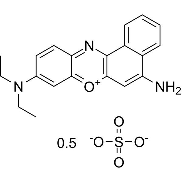
Nile Blue A sulfateamp;;(Synonyms: Nile blue sulfate)

Nile Blue A sulfate Chemical Structure

CAS No. : 3625-57-8

规格 价格 是否有货 数量
Free Sample (0.1-0.5 mg) ; Apply now ;
10;mM;*;1 mL in DMSO ¥550 In-stock
100 mg ¥500 In-stock
200 mg ; 询价 ;
500 mg ; 询价 ;

* Please select Quantity before adding items.

Nile Blue A sulfate 相关产品

bull;相关化合物库:

  • Bioactive Compound Library Plus

生物活性

Nile Blue A (Nile blue sulfate) is used to differentiate melanins and lipofuscins. It is also useful for staining fats and preparation of an amperometric glucose sensor[1].

体外研究
(In Vitro)

Nile blue A is a basic oxazine dye which is soluble in water and ethyl alcohol. Nile blue A is a satisfactory stain for PHB granules in bacteria and is in fact superior to Sudan black B for this purpose. Poly-p3-hydroxybutyrate granules exhibits a strong orange fluorescence when stained with Nile blue A. Nile blue A appears to stain many more PHB granules than Sudan black B does and is not as easily ished from the cell by decolorization procedures[1]. Nile blue A is used as a stain for polyhydroxyalkanoic acid-accumulating microorganisms or to detect polyhydroxyalkanoic acids in microorganisms. Escherichia coli cells that do not accumulate detectable polyhydroxyalkanoic acids can be stained with Nile blue A and that this staining is sufficient for identifying these cells in fluorescence-activated cell sorting (FACS) experiments. Nile blue A staining does not affect either surface display of peptides or specific labeling of these peptides by a second fluorescence. Staining E. coli for flow cytometry using Nile blue A is an easy-to-handle and low-cost alternative to other fluorescent dyes or the intracellular expression of, for example, green fluorescent protein[2]. Nile blue A is one of the most studied benzophenoxazine dyes, as a potent photosensitizer for photodynamic therapy. The dye when administered intravenously disperses throughout the body by circulating through blood and is taken up by most cells that emphasize its interaction with various biomolecule[3].

MCE has not independently confirmed the accuracy of these methods. They are for reference only.

分子量

366.42

Formula

C20H20N3O5S

CAS 号

3625-57-8

中文名称

硫酸耐尔蓝A;硫酸尼罗蓝

运输条件

Room temperature in continental US; may vary elsewhere.

储存方式

4deg;C, sealed storage, away from moisture and light

*In solvent : -80deg;C, 6 months; -20deg;C, 1 month (sealed storage, away from moisture and light)

溶解性数据
In Vitro:;

DMSO : ≥ 150 mg/mL (409.37 mM)

H2O : < 0.1 mg/mL (ultrasonic;warming) (insoluble)

* “≥” means soluble, but saturation unknown.

配制储备液
浓度 溶剂体积 质量 1 mg 5 mg 10 mg
1 mM 2.7291 mL 13.6455 mL 27.2911 mL
5 mM 0.5458 mL 2.7291 mL 5.4582 mL
10 mM 0.2729 mL 1.3646 mL 2.7291 mL

*

请根据产品在不同溶剂中的溶解度选择合适的溶剂配制储备液;一旦配成溶液,请分装保存,避免反复冻融造成的产品失效
储备液的保存方式和期限:-80°C, 6 months; -20°C, 1 month (sealed storage, away from moisture and light)。-80°C 储存时,请在 6 个月内使用,-20°C 储存时,请在 1 个月内使用。

参考文献
  • [1]. Ostle AG, et al. Nile blue A as a fluorescent stain for poly-beta-hydroxybutyrate. Appl Environ Microbiol. 1982 Jul;44(1):238-41.

    [2]. Betscheider D, et al. Nile blue A for staining Escherichia coli in flow cytometer experiments. Anal Biochem. 2009 Jan 1;384(1):194-6.

    [3]. Mishra SS, et al. Spectroscopic investigation of interaction of Nile Blue A, a potent photosensitizer, with bile salts in aqueous medium. J Photochem Photobiol B. 2014 Dec;141:67-75.

Cell Assay
[1][2][3]

1% aqueous solution of Nile blue A is prepared and filtered before use. Mild heating may be necessary to fully dissolve the stain. Heat-fixed smears of bacterial cells are stained with the Nile blue A solution at 55°C for 10 min in a coplin staining jar. After being stained, the slides are washed with tap water to remove excess stain and with 8% aqueous acetic acid for 1 min. The stained smear is washed and blotted dry with bibulous paper, remoistened with tap water, and covered with a no. 1 glass cover slip. The preparation is examined with a Nikon Labphot microscope with an episcopic fluorescence attachment[1]. The PHA− strain Escherichia coli UT5600(DE3) is stained with Nile blue A. In an Erlenmeyer flask, 20 mL of Luria–Bertani (LB) broth is inoculated with one colony of UT5600(DE3) and incubated for 14 h at 37 °C and 200 rpm. Subsequently, 20 mL of LB broth containing Nile blue A in a final concentration of 0.5 μg/mL is inoculated with 200 μL of the 14-h culture and cultured to an optical density at 578 nm (OD578) of 0.6. As a control, 20 mL of LB broth (without Nile blue A) is inoculated with 200 μL of the 14-h culture and is also cultured to an optical density at OD578 of 0.6. Every 20 min, the OD578 is determined for both cultures to verify whether there is any influence of the dye on the growth of the bacteria[2]. Nile blue A stock solution is prepared using ethanol as the solvent. The concentration of NB is maintained at 5 μM for all the studies. The solutions are left for 1 h to achieve equilibrium before spectral measurements. The absorption spectra are recorded using Shimadzu Spectrophotometer (UV-1800) and the emission spectra are recorded using Jobin–Yvon Spectrofluorimeter. A 450 nm nano-LED is used as the light source and the fluorescence lifetime is collected at λem=672 nm[3].

MCE has not independently confirmed the accuracy of these methods. They are for reference only.

参考文献
  • [1]. Ostle AG, et al. Nile blue A as a fluorescent stain for poly-beta-hydroxybutyrate. Appl Environ Microbiol. 1982 Jul;44(1):238-41.

    [2]. Betscheider D, et al. Nile blue A for staining Escherichia coli in flow cytometer experiments. Anal Biochem. 2009 Jan 1;384(1):194-6.

    [3]. Mishra SS, et al. Spectroscopic investigation of interaction of Nile Blue A, a potent photosensitizer, with bile salts in aqueous medium. J Photochem Photobiol B. 2014 Dec;141:67-75.

尼罗红/油红O 脂肪染色试剂 Nile Red/Oil Red O

尼罗红/油红O 脂肪染色试剂
Nile Red/Oil Red O

  • 产品特性
  • 相关资料
  • Q&A
  • 参考文献

尼罗红/油红O 脂肪染色试剂                              Nile Red/Oil Red O尼罗红·油红O

脂肪染色试剂


◆特点


● 能使用荧光显微镜以及流式细胞仪进行细胞内脂肪滴检测的活体染色试剂。

● 可简便快捷定量检测细胞内中性脂质等脂质的局部定位。


尼罗红/油红O 脂肪染色试剂                              Nile Red/Oil Red O

油红O

案例


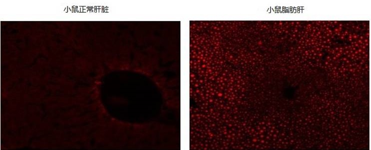
1、尼罗红:小鼠肝脏染色案例


尼罗红/油红O 脂肪染色试剂                              Nile Red/Oil Red O


观察到脂肪肝中小叶周边脂肪的堆积

图片提供:大阪保健医疗大学研究院  柴田雅朗,国立循环器官疾病研究中心 斯波真理子


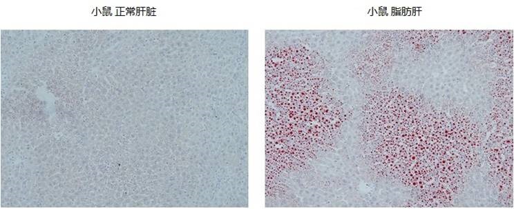
2、油红O:小鼠肝脏 染色案例


尼罗红/油红O 脂肪染色试剂                              Nile Red/Oil Red O


观察到脂肪肝中小叶外周性脂肪堆积

欲了解使用方法案例请看相关资料

◆使用方法案例


● 油红O

脂肪(中性脂肪)—橙红色~深红色

保存液—在100 mL 99%异丙醇添加0.3 g油红O,密闭容器60℃,放置过夜,制作饱和溶液。

使用液—保存液:蒸馏水以6:4混合,充分搅拌,10~30分钟后,过滤制成染色液。


● 尼罗蓝—硫酸氢盐

可同时分别染色酸性脂质和中性脂质。

酸性脂质—蓝色、中性脂质—红色

染色液—向100 mL蒸馏水中溶解7 g尼罗蓝—硫酸氢盐粉末,60℃或者37℃,放置过夜。

制成饱和溶液。然后冷却至室温,使用前请过滤。


● 苏丹Ⅲ

脂肪(中性脂肪)—橙黄色~橙红色

染色液—向100 mL 70%酒精溶解2 g苏丹Ⅲ,密闭容器60℃,放置过夜,制作饱和溶液。然后冷却至室温,过滤保存。


● 苏丹Ⅳ

苯环添加2个甲基,脂溶性增强,比苏丹Ⅲ的染色效果更稳定。

脂肪(中性脂肪)—橙黄色~橙红色

染色液—向100 mL 70%酒精添加苏丹Ⅳ 2 g,密闭容器60℃,放置一夜,制作饱和溶液。然后在室温冷却,过滤保存。


● 苏丹黑B

和其他苏丹染色比较,染色性稳定。

脂肪—黑色~蓝黑色

染色液—向100 mL 70%酒精添加0.1 g苏丹黑B,60℃加热溶解。

冷却至室温,过滤后使用。2周内使用。


产品列表
产品编号 产品名称 产品规格 产品等级 备注
144-08811 尼罗红
 Nile Red
25 mg 病理研究用
140-08813 尼罗红
 Nile Red
100 mg 病理研究用
154-02072 油红O
 Oil Red O
25 g 病理研究用
141-06822 尼罗蓝—硫酸氢盐
 Nile Blue Hydrogensulfate
25 g 病理研究用
192-04392 苏丹Ⅲ
 Sudan III
25 g 和光特级
194-07652 苏丹红IV
 Sudan IV
25 g 和光一级
192-04412 苏丹黑B
 Sudan Black B
25 g 和光一级

Nile Red 尼罗红 品牌:FUJIFILM Wako


Nile Red

尼罗红

品牌:FUJIFILM Wako
CAS No.:7385-67-3
储存条件:25℃以下
纯度:
产品编号

(生产商编号)

等级 规格 运输包装 零售价(RMB) 库存情况 参考值

140-08813

for Pathology Research 100mg 1,850.00


* 干冰运输、大包装及大批量的产品需酌情添加运输费用


* 零售价、促销产品折扣、运输费用、库存情况、产品及包装规格可能因各种原因有所变动,恕不另行通知,确切详情请联系上海金畔生物科技有限公司。

Nile Red 尼罗红 品牌:FUJIFILM Wako


Nile Red

尼罗红

品牌:FUJIFILM Wako
CAS No.:7385-67-3
储存条件:25℃以下
纯度:
产品编号

(生产商编号)

等级 规格 运输包装 零售价(RMB) 库存情况 参考值

144-08811

for Pathology Research 25mg 560.00


* 干冰运输、大包装及大批量的产品需酌情添加运输费用


* 零售价、促销产品折扣、运输费用、库存情况、产品及包装规格可能因各种原因有所变动,恕不另行通知,确切详情请联系上海金畔生物科技有限公司。

Nile Blue Hydrogensulfate 尼罗蓝硫酸盐 品牌:FUJIFILM Wako


Nile Blue Hydrogensulfate

尼罗蓝硫酸盐

品牌:FUJIFILM Wako
CAS No.:3625-57-8
储存条件:室温
纯度:
产品编号

(生产商编号)

等级 规格 运输包装 零售价(RMB) 库存情况 参考值

141-06822

for Pathology Research 25 g

Nile Blue Hydrogensulfate                                                      尼罗蓝硫酸盐            品牌:FUJIFILM Wako


* 干冰运输、大包装及大批量的产品需酌情添加运输费用


* 零售价、促销产品折扣、运输费用、库存情况、产品及包装规格可能因各种原因有所变动,恕不另行通知,确切详情请联系上海金畔生物科技有限公司。

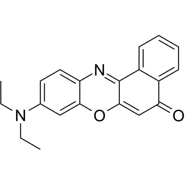
Nile Red(Synonyms: 尼罗红 Nile Blue A oxazone Phenoxazone 9)

Nile Red;(Synonyms: 尼罗红; Nile Blue A oxazone; Phenoxazone 9) 纯度: 98.02%

Nile Red (Nile Blue A oxazone) 是一种针对细胞内脂质小滴和中性脂质的选择性疏水性荧光染料。Nile Red 在所有有机溶剂中均具有强烈的荧光性,其荧光颜色范围从金黄色到深红色。

Nile Redamp;;(Synonyms: 尼罗红; Nile Blue A oxazone;  Phenoxazone 9)

Nile Red Chemical Structure

CAS No. : 7385-67-3

规格 价格 是否有货 数量
Free Sample (0.1-0.5 mg) ; Apply now ;
10;mM;*;1 mL in DMSO ¥440 In-stock
10 mg ¥400 In-stock
50 mg ¥650 In-stock
100 mg ¥800 In-stock
500 mg ¥1400 In-stock
1 g ; 询价 ;
5 g ; 询价 ;

* Please select Quantity before adding items.

Nile Red 相关产品

bull;相关化合物库:

  • Bioactive Compound Library Plus

生物活性

Nile Red (Nile Blue A oxazone) is a selective and hydrophobic fluorescent stain for intracellular lipid droplets and neutral lipids. Nile Red is intensely fluorescent in all organic solvents and the fluorescence colors range from golden yellow to deep red[1][2].

体外研究
(In Vitro)

Nile red-stained, lipid droplet-filled macrophages exhibit greater fluorescence intensity than does Nile red-stained control macrophages, and the two cell populations could be differentiated and analyzed by flow cytofluorometry. Better selectivity for cytoplasmic lipid droplets is obtained when the cells are viewed for yellow-gold fluorescence (excitation, 450-500 nm; emission, greater than 528 nm) rather than red fluorescence (excitation, 515-560 nm; emission, greater than 590 nm)[1].
Nile red is strongly fluorescent, but only in the presence of a hydrophobic environment. Nile red is very soluble in the lipids it is intended to show, and it does not interact with any tissue constituent except by solution[1].
Spectral and physicochemical properties of the lipophilic dye Nile red induce a yellow-gold-spectral shift in its excitation-emission peak, allowing it to fluoresce in the green emission spectrum only when in a lipid-rich environment, but not in more polar environments[4].

MCE has not independently confirmed the accuracy of these methods. They are for reference only.

体内研究
(In Vivo)

When Nile red-stained Caenorhabditis elegans is viewed for green fluorescence, discrete lipid bodies can be observed throughout the intestine and other tissues either in clusters or evenly dispersed, depending on the animal’s genotype or experimental treatment[3].

MCE has not independently confirmed the accuracy of these methods. They are for reference only.

分子量

318.37

Formula

C20H18N2O2

CAS 号

7385-67-3

中文名称

尼罗红

运输条件

Room temperature in continental US; may vary elsewhere.

储存方式

4deg;C, protect from light

*In solvent : -80deg;C, 6 months; -20deg;C, 1 month (protect from light)

溶解性数据
In Vitro:;

DMSO : ≥ 50 mg/mL (157.05 mM)

H2O : 0.1 mg/mL (0.31 mM; Need ultrasonic)

* “≥” means soluble, but saturation unknown.

配制储备液
浓度 溶剂体积 质量 1 mg 5 mg 10 mg
1 mM 3.1410 mL 15.7050 mL 31.4100 mL
5 mM 0.6282 mL 3.1410 mL 6.2820 mL
10 mM 0.3141 mL 1.5705 mL 3.1410 mL

*

请根据产品在不同溶剂中的溶解度选择合适的溶剂配制储备液;一旦配成溶液,请分装保存,避免反复冻融造成的产品失效
储备液的保存方式和期限:-80°C, 6 months; -20°C, 1 month (protect from light)。-80°C 储存时,请在 6 个月内使用,-20°C 储存时,请在 1 个月内使用。

参考文献
  • [1]. Greenspan P, et al. Nile red: a selective fluorescent stain for intracellular lipid droplets. J Cell Biol. 1985 Mar;100(3):965-73.

    [2]. Gibrán S Alemán-Nava, et al. How to use Nile Red, a selective fluorescent stain for microalgal neutral lipids. J Microbiol Methods. 2016 Sep;128:74-79.

    [3]. Wilber Escorcia, et al. Quantification of Lipid Abundance and Evaluation of Lipid Distribution in Caenorhabditis elegans by Nile Red and Oil Red O Staining. J Vis Exp. 2018 Mar 5;(133):57352.

    [4]. Elizabeth C Pino, et al. Biochemical and high throughput microscopic assessment of fat mass in Caenorhabditis elegans. J Vis Exp. 2013 Mar 30;(73):50180.