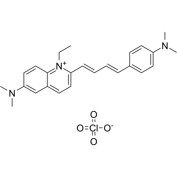
LDS-751; 纯度: 99.30%
LDS-751 是一种主要检测 DNA 的核酸染色剂。LDS-751 是一种主要检测DNA的核酸染色剂。LDS-751 对 DNA 有很高的亲和力,结合后荧光增强,但最大发射波长为 670nm。LDS-751 和 Thiazole orange 可用于红细胞、血小板、网织红细胞和有核细胞的分化,并能在 488nm 处激发。

LDS-751 Chemical Structure
CAS No. : 181885-68-7
| 规格 | 价格 | 是否有货 | 数量 |
|---|---|---|---|
| 5 mg | ¥1950 | In-stock | |
| 10 mg | ; | 询价 | ; |
| 50 mg | ; | 询价 | ; |
* Please select Quantity before adding items.
LDS-751 相关产品
bull;相关化合物库:
- Bioactive Compound Library Plus
| 生物活性 |
LDS-751 is a nucleic acid stain principally detecting DNA. LDS-751 has high affinity for DNA and undergoes fluorescence enhancement upon binding, but with maximal emission at 670 nm. LDS-751 and Thiazole orange are used to differentiate erythrocytes, platelets, reticulocytes, and nucleated cells, and both can be excited at 488 nm[1][2][3]. |
||||||||||||||||
|---|---|---|---|---|---|---|---|---|---|---|---|---|---|---|---|---|---|
| 体外研究 (In Vitro) |
The LDS-751 permits the discrimination of intact cells from residual erythrocyte ghosts, platelets and damaged nucleated cells. The forward and orthogonal fight scattering signals of the intact cells, identified by LDS-751 staining allow a clear separation between the major leukocyte populations since the damaged nucleated cells, erythrocytes, erythrocyte cell ghosts and platelets are removed from the display[1]. MCE has not independently confirmed the accuracy of these methods. They are for reference only. |
||||||||||||||||
| 分子量 |
471.98 |
||||||||||||||||
| Formula |
C25H30ClN3O4 |
||||||||||||||||
| CAS 号 |
181885-68-7 |
||||||||||||||||
| 运输条件 |
Room temperature in continental US; may vary elsewhere. |
||||||||||||||||
| 储存方式 |
-20deg;C, sealed storage, away from moisture and light *该产品在溶液状态不稳定,建议您现用现配,即刻使用。 |
||||||||||||||||
| 溶解性数据 |
In Vitro:;
DMSO : 83.33 mg/mL (176.55 mM; Need ultrasonic) 配制储备液
*
请根据产品在不同溶剂中的溶解度,选择合适的溶剂配制储备液;该产品在溶液状态不稳定,建议您现用现配,即刻使用。 In Vivo:
请根据您的实验动物和给药方式选择适当的溶解方案。以下溶解方案都请先按照 In Vitro 方式配制澄清的储备液,再依次添加助溶剂: ——为保证实验结果的可靠性,澄清的储备液可以根据储存条件,适当保存;体内实验的工作液,建议您现用现配,当天使用; 以下溶剂前显示的百
|
||||||||||||||||
| 参考文献 |
|
| Cell Assay |
Blood samples (1 mL) are obtained using a sterile Butterfly-21 needle and plastic syringe from the antecubital vein of normal healthy volunteers who have given their informed consent. They are immediately transferred to plastic tubes containing 17.3 mg of phenylmethylsulphonyl fluoride (PMSF) and 1 mL of LDS-751 at room temperature. Aliquots (25 μL) are incubated for 5 min with between 2 μL and 5 μL of undiluted monoclonal antibodies, then diluted with 0.5 mL of 1% BSA in Hepesbuffered Hanks’ balanced salts solution (HHBSS) and examined by flow cytometry[2]. MCE has not independently confirmed the accuracy of these methods. They are for reference only. |
|---|---|
| 参考文献 |
|
