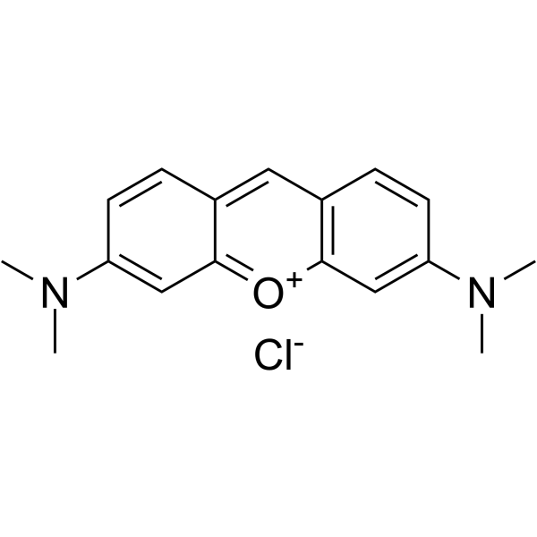
Pyronin Y;(Synonyms: 派洛宁Y; Pyronine G; C.I. 45005)
Pyronin Y (Pyronine G) 是一种可插入 RNA 的阳离子染料,可以靶向包括 RNA,DNA 和细胞器的细胞结构。Pyronin Y 与双链核酸(尤其是 RNA) 形成荧光复合物,可以对细胞 RNA 进行半定量分析。Pyronin Y 可用于鉴定活细胞中核糖核蛋白复合物的特定 RNA 亚种。

Pyronin Y Chemical Structure
CAS No. : 92-32-0
| 规格 | 价格 | 是否有货 | 数量 |
|---|---|---|---|
| 10;mM;*;1 mL in DMSO | ¥660 | In-stock | |
| 100 mg | ¥500 | In-stock | |
| 200 mg | ; | 询价 | ; |
| 500 mg | ; | 询价 | ; |
* Please select Quantity before adding items.
Pyronin Y 相关产品
bull;相关化合物库:
- Bioactive Compound Library Plus
| 生物活性 |
Pyronin Y (Pyronine G) is a cationic dye that intercalates RNA and has been used to target cell structures including RNA, DNA and organelles. Pyronin Y forms fluorescent complexes with double-stranded nucleic acids (especially RNA) enabling semi-quantitative analysis of cellular RNA. Pyronin Y can be used to identify specific RNA subspecies of ribonuclear proteins complexes in live cells[1][2][5]. |
||||||||||||||||
|---|---|---|---|---|---|---|---|---|---|---|---|---|---|---|---|---|---|
| 体外研究 (In Vitro) |
Pyronin Y forms fluorescent complexes with double-stranded nucleic acids, especially RNA, enabling semi-quantitative analysis of cellular RNA in flow cytometry, to estimate the RNA content per cell in formalin fixed EL4 leukosis tumor cells, enzyme dispersed R3327-G rat prostatic adenocarcinoma cells, mouse spleen cells stimulated with concanavalin A, and human peripheral blood lymphocytes stimulated with phytohemagglutinin[1]. MCE has not independently confirmed the accuracy of these methods. They are for reference only. |
||||||||||||||||
| 分子量 |
302.80 |
||||||||||||||||
| Formula |
C17H19ClN2O |
||||||||||||||||
| CAS 号 |
92-32-0 |
||||||||||||||||
| 中文名称 |
派洛宁Y;吡罗红G |
||||||||||||||||
| 运输条件 |
Room temperature in continental US; may vary elsewhere. |
||||||||||||||||
| 储存方式 |
-20deg;C, sealed storage, away from moisture and light *In solvent : -80deg;C, 6 months; -20deg;C, 1 month (sealed storage, away from moisture and light) |
||||||||||||||||
| 溶解性数据 |
In Vitro:;
DMSO : 25 mg/mL (82.56 mM; Need ultrasonic) H2O : 4 mg/mL (13.21 mM; Need ultrasonic) 配制储备液
*
请根据产品在不同溶剂中的溶解度选择合适的溶剂配制储备液;一旦配成溶液,请分装保存,避免反复冻融造成的产品失效。 |
||||||||||||||||
| 参考文献 |
|
| Cell Assay [2] |
The cells are resuspended and stained for 30 min in 1.0 mL of 0.01% pyronin Y in sodiumacetate buffer (1.0 M, pH 4.7). For this purpose 1.0 g of pyronin Y is dissolved in 100 mL distilled water and this solution is purified by chloroform extraction (three fractions of 100 mL). The purified pyronin Y solution is diluted with 1.0 M sodiumacetate buffer pH 4.7 to the appropriate concentration and filtered through paper filters before use. Stained cell samples are studied by fluorescence microscopy (excitation filters SP 560 and LP 515, chromatic beam splitter at 580 nm, barrier filter LP 580) or by flow cytometry[2]. MCE has not independently confirmed the accuracy of these methods. They are for reference only. |
|---|---|
| 参考文献 |
|
