SP6 RNA Polymerase, recombinant, Solution 品牌:FUJIFILM Wako


SP6 RNA Polymerase, recombinant, Solution

品牌:FUJIFILM Wako
CAS No.:
储存条件:-20℃
纯度:
产品编号

(生产商编号)

等级 规格 运输包装 零售价(RMB) 库存情况 参考值

298-69101

for Genetic Research 5000 units

SP6 RNA Polymerase, recombinant, Solution                                                                品牌:FUJIFILM Wako


* 干冰运输、大包装及大批量的产品需酌情添加运输费用


* 零售价、促销产品折扣、运输费用、库存情况、产品及包装规格可能因各种原因有所变动,恕不另行通知,确切详情请联系上海金畔生物科技有限公司。

RNA Ligase, T4 pahge, recombinant, Soln. RNA连接酶 品牌:FUJIFILM Wako


RNA Ligase, T4 pahge, recombinant, Soln.

RNA连接酶

品牌:FUJIFILM Wako
CAS No.:9015-85-4
储存条件:-20℃
纯度:
产品编号

(生产商编号)

等级 规格 运输包装 零售价(RMB) 库存情况 参考值

189-01501

for Genetic Research 1000 U

RNA Ligase, T4 pahge, recombinant, Soln.                                                      RNA连接酶            品牌:FUJIFILM Wako


* 干冰运输、大包装及大批量的产品需酌情添加运输费用


* 零售价、促销产品折扣、运输费用、库存情况、产品及包装规格可能因各种原因有所变动,恕不另行通知,确切详情请联系上海金畔生物科技有限公司。

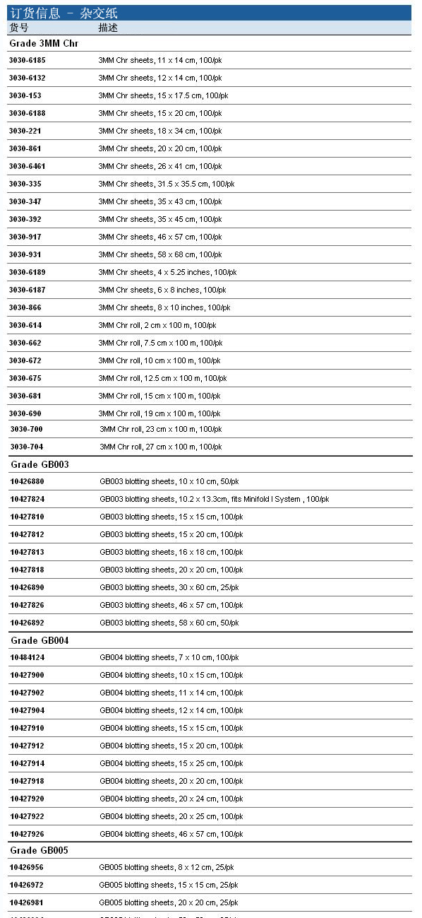
3030-861-英国whatman层析纸3MM杂交用纸

  • 型号 3030-861
  • 品牌 GE whatman沃特曼
  • 【简单介绍】
    品牌 其他品牌

    英国whatman层析纸3MM杂交用纸
    【实验目的】
    掌握RNA印迹技术的基本原理,学习RNA印迹技术的操作方法,了解RNA印迹技术的意义与应用。

    英国whatman层析纸3MM杂交用纸 3030-861

    【实验目的】

    掌握RNA印迹技术的基本原理,学习RNA印迹技术的操作方法,了解RNA印迹技术的意义与应用。

    【实验原理】

    将RNA变性及电泳分离后,转移到固相支持物上,用于与DNA探针杂交以鉴定其中特定RNA分子的大小与含量。基本原理与Southern印迹相同,但RNA变性方法与DNA不同,不能用碱变性,因为碱会导致RNA的水解。

    Northern印迹主要用于组织细胞靶基因表达水平的研究以及对同一组织细胞的不同基因间的表达水平进行比较,或者对不同组织细胞间相同基因的表达水平进行比较。

    【实验对象】

    待测RNA样品。

    【试剂与器材】

    1. 焦碳酸二乙酯 (DEPC)。

    2. 甲醛变性琼脂糖凝胶电泳所需试剂。

    3. 0.5 mol / L乙酸铵(NH4Ac)。

    4. 溴化乙啶 (EB):0.5μg/ml,溶于0.5mol/L乙酸铵中。

    5. 0.05mol / L NaOH/1.5mol/L NaCI(选用)。

    6. 0.5mol / L Tris·CI (pH 7.4) /1.5mol/L NaCl(选用)。

    7. 20×SSC、6×SSC和2×SSC。

    8. 亚甲蓝(终浓度为0.03%),溶于0.3mol / L (pH 5.2)的乙酸钠缓冲液中 (选用)。

    9. *纤维素 (NC) 膜或尼龙膜。

    【实验方法与步骤】

    1. RNA甲醛变性凝胶电泳(5V/cm, 3h),(见附录三)。

    电泳结束后用EB染色:取出凝胶,切下需要染色的泳道,放置于无RNA酶的玻璃平皿,加入0.5mol /L乙酸铵浸泡20min。换液再浸泡20min以除去甲醛,将凝胶转入含0.5(g / ml EB的0.5mol /L乙酸铵溶液中染色40min,必要时,以0.5mol / L乙酸铵脱色1h。

    3.在紫外透射仪中使凝胶中的RNA显迹,沿凝胶的边缘放一把尺子拍照,以便随后能确定膜中的条带。

    4.将非染色的部分凝胶放置于无RNA酶的玻璃平皿中,加足够的DEPC处理的去离子水浸洗几次以除去甲醛。

    5. 凝胶浸泡在10倍体积的0.05mol/L NaOH / 1.5mol/LNaCl中30min,使RNA部分水解。接着浸泡于10倍体积的0.5mol/L Tris·C1(pH 7.4) / 1.5mol/LNaCl缓冲液中和30min (可选), 将胶的多余部分切除,并切边标记。

    6. 将溶液换成10倍体积的20×SSC,浸泡45min。

    7. 在一玻璃或塑料平皿中放置-块比凝胶略大的有机玻璃作为平台(必要时,可并排放两块或更多) ,加入足够的20×SSC以使平台半淹没在液体中。

    8. 构筑转移平台,剪3张与平台同样大小的Whatman 3MM滤纸,放在平台表面,以20×SSC润湿。把凝胶置于滤纸表面,用玻棒滚动表面以压挤去除气泡。切4条塑料保鲜膜覆盖在凝胶的边缘。

    9.剪一片与凝胶暴露面积大小相当的尼龙膜或NC膜,在一个无RNA酶的玻璃平皿中加入约0.5mm深的用DEPC处理的去离子水,将膜漂在水面使之润湿,直至膜沉没在水中。如果是尼龙膜,只需让膜在水中泡上5min,如果是NC膜,则换成20×SSC再浸泡10min。

    10.将润湿的膜放在凝胶表面,用干净刀片切去滤膜一角,使其与凝胶的切角相对应,排去气泡,以20×SSC漂洗膜表面。切 3张与膜大小相当的湿润的Whatman3MM滤纸,放于膜的表面。赶出气泡。然后将与膜大小相当的纸巾整齐地堆放在滤纸的表面,高约4cm。

    11. 在纸堆的顶部放一块玻璃平板,压一重物 (0.2~0.4kg),放置12h左右。

    12.拆卸转移装置,回收膜和压扁了的凝胶。在膜上用铅笔标上加样孔的位置和方向。


    13.用2×SSC洗膜,然后放于滤纸上,让膜于室温干燥30分钟。NC膜则应夹于2张Whatman 3MM滤纸之间,80℃真空干烤2h。将干的转印膜置于可透过紫外线的塑料保鲜膜中,带有RNA的一面朝下,置于紫外透射仪(波长在254nm)上,以合适的照射强度进行紫外线交联,以固定RNA。备作核酸杂交。

    14. 凝胶可按步骤2,用EB染色以检查转印效率。

    【注意事项】

    1.为了抑制RNA酶的活性,所有用于Northern印迹的溶液,均须用经DEPC处理过的无菌去离子水配制。DEPC是一种致癌剂,须小心操作。尤其在用DEPC处理乙酸铵时,因为DEPC能与铵离子反应产生氨基甲酸乙酯(一种潜在的致癌剂),故在以DEPC处理乙酸铵时,更须分外小心。

    2.RNA极易被环境中存在的RNA酶降解,因此须特别警惕RNA酶的污染

     用途:本品仅供科研,不得用于其它用途。

    英国whatman层析纸3MM杂交用纸 3030-861 订货信息:

    3030-861-英国whatman层析纸3MM杂交用纸

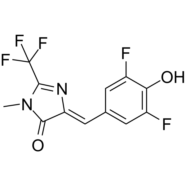
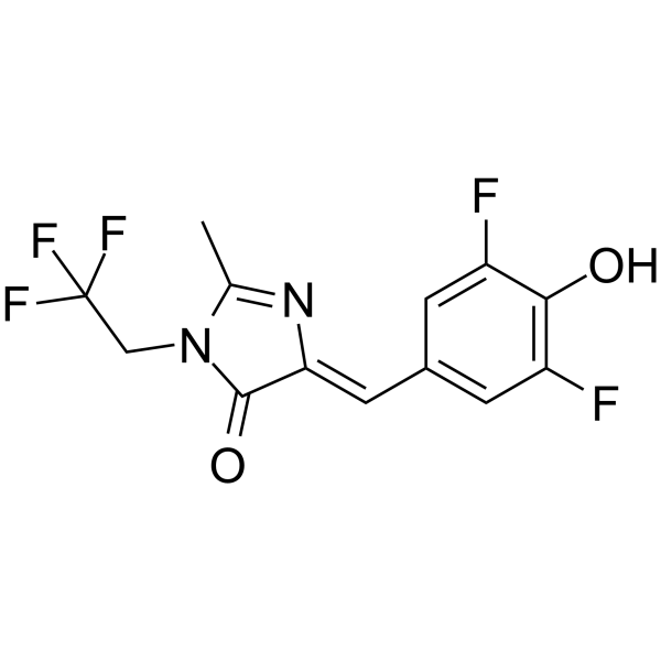
荧光染料DFHBI-2T

荧光染料Dye Reagents DFHBI-2T;

DFHBI-2T 是一种可透过膜的 RNA 适配体激活的荧光探针 (ex/em=500 nm/523 nm)。DFHBI-2T 可以用于活细胞中的 RNA 成像。

DFHBI-2T

DFHBI-2T Chemical Structure

CAS No. : 1539318-40-5

规格 价格 是否有货
5 mg ¥4800 询问价格 货期
10 mg ¥8000 询问价格 货期
25 mg ¥15500 询问价格 货期

* Please select Quantity before adding items.

生物活性

DFHBI-2T is a membrane-permeable RNA aptamers-activated fluorescence probe (ex/em=500 nm/523 nm). DFHBI-2T is used to image RNA in live cells[1][2].

体外研究
(In Vitro)

Spinach2 imaged DFHBI-2T results in fluorescence that is detectable using the yellow emission channel[2].

MCE has not independently confirmed the accuracy of these methods. They are for reference only.

分子量

306.19

Formula

C12H7F5N2O2

CAS 号

1539318-40-5

运输条件

Room temperature in continental US; may vary elsewhere.

储存方式

Please store the product under the recommended conditions in the Certificate of Analysis.

参考文献
  • [1]. Wenjiao Song, et al. Plug-and-play fluorophores extend the spectral properties of Spinach. J Am Chem Soc. 2014 Jan 29;136(4):1198-201.

    [2]. Robert J Trachman 3rd, et al. Structural Principles of Fluorescent RNA Aptamers. Trends Pharmacol Sci. 2017 Oct;38(10):928-939.

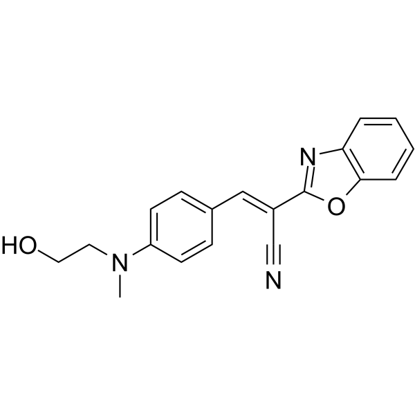
荧光染料HBC525

荧光染料Dye Reagents HBC525;

HBC525 是一种类 HBC 荧光团和荧光 RNA 适体 (Kd=3.8 nM)。HBC525 可以直接作为融合标签用于感兴趣的细胞 RNA 的成像和跟踪。荧光 RNA 适体也被用于构建各种有趣的用于细胞靶点检测和成像的动态 RNA 纳米工具。

HBC525

HBC525 Chemical Structure

CAS No. : 2174014-64-1

规格 是否有货
100 mg ; 询价 ;
250 mg ; 询价 ;
500 mg ; 询价 ;

* Please select Quantity before adding items.

生物活性

HBC525 is a HBC-like fluorophore and a fluorogenic RNA aptamer (Kd=3.8 nM). HBC525 can be directly used as fusion tags for the imaging and tracking of cellular RNAs of interest. Fluorogenic RNA aptamers have also been used to construct various interesting dynamic RNA nanodevices for cellular target detection and imaging[1][2].

IC50 Target

HBC525: 491/525 nm (Ex/Em)[2]

体内研究
(In Vivo)

荧光RNA适体

MCE has not independently confirmed the accuracy of these methods. They are for reference only.

分子量

319.36

Formula

C19H17N3O2

CAS 号

2174014-64-1

运输条件

Room temperature in continental US; may vary elsewhere.

储存方式

Please store the product under the recommended conditions in the Certificate of Analysis.

参考文献
  • [1]. Chen X, et al. Visualizing RNA dynamics in live cells with bright and stable fluorescent RNAs. Nat Biotechnol. 2019;37(11):1287-1293.

    [2]. Yu Q, et al. Genetically encoded RNA nanodevices for cellular imaging and regulation. Nanoscale. 2021;13(17):7988-8003.

DFHBI-1T

DFHBI-1T; 纯度: 98.82%

DFHBI-1T 是一种可透过膜的 RNA 适配体激活的荧光探针 (ex/em=472 nm/507 nm)。DFHBI-1T 与 RNA 适配体 (Spinach, Spinach2, iSpinach, Broccoli) 结合并产生特定的荧光和较低的背景荧光。DFHBI-1T 可以用于活细胞中的 RNA 成像。

DFHBI-1Tamp;;

DFHBI-1T Chemical Structure

CAS No. : 1539318-36-9

规格 价格 是否有货 数量
Free Sample (0.1-0.5 mg) ; Apply now ;
5 mg ¥1200 In-stock
10 mg ¥2000 In-stock
25 mg ¥4200 In-stock
50 mg ¥6500 In-stock
100 mg ¥9800 In-stock
200 mg ; 询价 ;
500 mg ; 询价 ;

* Please select Quantity before adding items.

DFHBI-1T 相关产品

bull;相关化合物库:

  • Bioactive Compound Library Plus

生物活性

DFHBI-1T is a membrane-permeable RNA aptamers-activated fluorescence probe (ex/em=472 nm/507 nm). DFHBI-1T binds to RNA aptamers (Spinach, Spinach2, iSpinach, and Broccoli) and causes specific fluorescence and lower background fluorescence. DFHBI-1T is used to image RNA in live cells[1][2].

体外研究
(In Vitro)

DFHBI-1T (20 μM; for 10 min) increases fluorescence in COS7 cells expressing (CGG)60-Spinach2 over DFHBI (20 μM)[1].
Broccoli-DFHBI-1T has ex/em=472 nm/507 nm and Spinach2-DFHBI-1T has ex/em=482 nm/505 nm[2].

MCE has not independently confirmed the accuracy of these methods. They are for reference only.

分子量

320.21

Formula

C13H9F5N2O2

CAS 号

1539318-36-9

运输条件

Room temperature in continental US; may vary elsewhere.

储存方式

-20deg;C, protect from light

*In solvent : -80deg;C, 6 months; -20deg;C, 1 month (protect from light)

溶解性数据
In Vitro:;

DMSO : 50 mg/mL (156.15 mM; Need ultrasonic)

配制储备液
浓度 溶剂体积 质量 1 mg 5 mg 10 mg
1 mM 3.1230 mL 15.6148 mL 31.2295 mL
5 mM 0.6246 mL 3.1230 mL 6.2459 mL
10 mM 0.3123 mL 1.5615 mL 3.1230 mL

*

请根据产品在不同溶剂中的溶解度选择合适的溶剂配制储备液;一旦配成溶液,请分装保存,避免反复冻融造成的产品失效
储备液的保存方式和期限:-80°C, 6 months; -20°C, 1 month (protect from light)。-80°C 储存时,请在 6 个月内使用,-20°C 储存时,请在 1 个月内使用。

In Vivo:

请根据您的实验动物和给药方式选择适当的溶解方案。以下溶解方案都请先按照 In Vitro 方式配制澄清的储备液,再依次添加助溶剂:

——为保证实验结果的可靠性,澄清的储备液可以根据储存条件,适当保存;体内实验的工作液,建议您现用现配,当天使用; 以下溶剂前显示的百
分比是指该溶剂在您配制终溶液中的体积占比;如在配制过程中出现沉淀、析出现象,可以通过加热和/或超声的方式助溶

  • 1.

    请依序添加每种溶剂:;10% DMSO ;; 40% PEG300 ;; 5% Tween-80 ;; 45% saline

    Solubility: 5.5 mg/mL (17.18 mM); Suspended solution; Need ultrasonic

    此方案可获得 5.5 mg/mL (17.18 mM) 的均匀悬浊液,悬浊液可用于口服和腹腔注射。

    以 1 mL 工作液为例,取 100 μL 55.0 mg/mL 的澄清 DMSO 储备液加到 400 μL PEG300 中,混合均匀;向上述体系中加入50 μL Tween-80,混合均匀;然后继续加入 450 μL生理盐水定容至 1 mL。

    将 0.9 g 氯化钠,完全溶解于 100 mL ddH₂O 中,得到澄清透明的生理盐水溶液

*以上所有助溶剂都可在 MCE 网站选购。
参考文献
  • [1]. Wenjiao Song, et al. Plug-and-play fluorophores extend the spectral properties of Spinach. J Am Chem Soc. 2014 Jan 29;136(4):1198-201.

    [2]. Grigory S Filonov, et al. Broccoli: rapid selection of an RNA mimic of green fluorescent protein by fluorescence-based selection and directed evolution. J Am Chem Soc. 2014 Nov 19;136(46):16299-308.

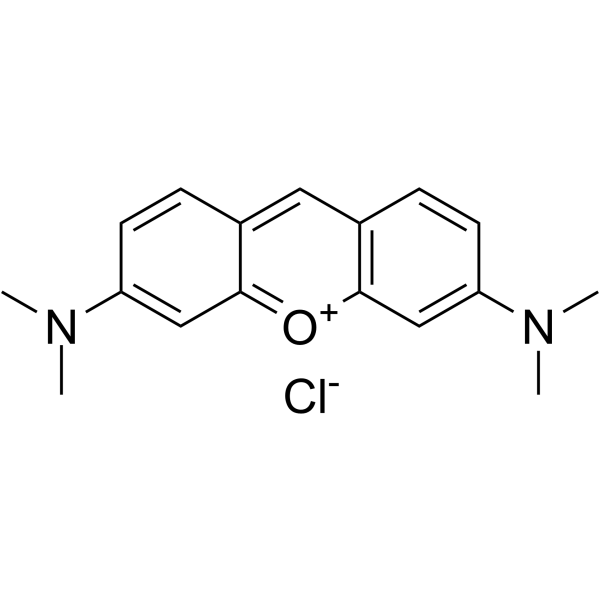
Pyronin Y(Synonyms: 派洛宁Y Pyronine G C.I. 45005)

Pyronin Y;(Synonyms: 派洛宁Y; Pyronine G; C.I. 45005)

Pyronin Y (Pyronine G) 是一种可插入 RNA 的阳离子染料,可以靶向包括 RNA,DNA 和细胞器的细胞结构。Pyronin Y 与双链核酸(尤其是 RNA) 形成荧光复合物,可以对细胞 RNA 进行半定量分析。Pyronin Y 可用于鉴定活细胞中核糖核蛋白复合物的特定 RNA 亚种。

Pyronin Yamp;;(Synonyms: 派洛宁Y; Pyronine G;  C.I. 45005)

Pyronin Y Chemical Structure

CAS No. : 92-32-0

规格 价格 是否有货 数量
10;mM;*;1 mL in DMSO ¥660 In-stock
100 mg ¥500 In-stock
200 mg ; 询价 ;
500 mg ; 询价 ;

* Please select Quantity before adding items.

Pyronin Y 相关产品

bull;相关化合物库:

  • Bioactive Compound Library Plus

生物活性

Pyronin Y (Pyronine G) is a cationic dye that intercalates RNA and has been used to target cell structures including RNA, DNA and organelles. Pyronin Y forms fluorescent complexes with double-stranded nucleic acids (especially RNA) enabling semi-quantitative analysis of cellular RNA. Pyronin Y can be used to identify specific RNA subspecies of ribonuclear proteins complexes in live cells[1][2][5].

体外研究
(In Vitro)

Pyronin Y forms fluorescent complexes with double-stranded nucleic acids, especially RNA, enabling semi-quantitative analysis of cellular RNA in flow cytometry, to estimate the RNA content per cell in formalin fixed EL4 leukosis tumor cells, enzyme dispersed R3327-G rat prostatic adenocarcinoma cells, mouse spleen cells stimulated with concanavalin A, and human peripheral blood lymphocytes stimulated with phytohemagglutinin[1].
A fluorescent staining procedure based on pyronin Y is described. The technique has been used to stain RNA in human reticulocytes for subsequent flow analysis and sorting[2].
In viable cells this dye also accumulates in mitochondria. At a concentration of 1.7 to 3.3 μM, pyronin Y is localized almost exclusively in mitochondria of cultured cells, similar to another mitochondria! probe, rhodamine 123. At that concentration PY is not toxic but suppressed cell growth, arresting cells[3].
Pyronin Y has long been used, in combination with other dyes such as Methyl Green, as a differential stain for nucleic acids in paraffin tissue sections. In sections stained with Methyl Green-pyronin Y, red blood cells, elastic fibre of blood vessels, zymogen granules of pancreatic acinar cells, surface membrane of heptocytes and kidney tubular cells showed strikingly strong green and/or red fluorescence, while the nuclei of cells appeared non-fluorescent[4].
Pyronin Y binds to double stranded RNA (dsRNA) including messenger RNA (mRNA), transfer RNA (tRNA) and ribosomal RNA (rRNA)[5].

MCE has not independently confirmed the accuracy of these methods. They are for reference only.

分子量

302.80

Formula

C17H19ClN2O

CAS 号

92-32-0

中文名称

派洛宁Y;吡罗红G

运输条件

Room temperature in continental US; may vary elsewhere.

储存方式

-20deg;C, sealed storage, away from moisture and light

*In solvent : -80deg;C, 6 months; -20deg;C, 1 month (sealed storage, away from moisture and light)

溶解性数据
In Vitro:;

DMSO : 25 mg/mL (82.56 mM; Need ultrasonic)

H2O : 4 mg/mL (13.21 mM; Need ultrasonic)

配制储备液
浓度 溶剂体积 质量 1 mg 5 mg 10 mg
1 mM 3.3025 mL 16.5126 mL 33.0251 mL
5 mM 0.6605 mL 3.3025 mL 6.6050 mL
10 mM 0.3303 mL 1.6513 mL 3.3025 mL

*

请根据产品在不同溶剂中的溶解度选择合适的溶剂配制储备液;一旦配成溶液,请分装保存,避免反复冻融造成的产品失效
储备液的保存方式和期限:-80°C, 6 months; -20°C, 1 month (sealed storage, away from moisture and light)。-80°C 储存时,请在 6 个月内使用,-20°C 储存时,请在 1 个月内使用。

参考文献
  • [1]. Pollack A, et al. Flow cytometric analysis of RNA content in different cell populations using pyronin Yand methyl green. Cytometry. 1982 Jul;3(1):28-35.

    [2]. Tanke HJ, et al. Flow cytometry of human reticulocytes based on RNA fluorescence. Cytometry. 1981 Mar;1(5):313-20.

    [3]. Darzynkiewicz Z, et al. Cytostatic and cytotoxic properties of pyronin Y: relation to mitochondrial localization of the dye and its interaction with RNA. Cancer Res. 1986 Nov;46(11):5760-6.

    [4]. Li B, et al. Pyronin Y as a fluorescent stain for paraffin sections. Histochem J. 2002 Jun-Jul;34(6-7):299-303.

    [5]. Laura M Andrews, et al. Spectral phasor analysis of Pyronin Y labeled RNA microenvironments in living cells. Biomed Opt Express. 2013 Jan 1;4(1):171-7.

Cell Assay
[2]

The cells are resuspended and stained for 30 min in 1.0 mL of 0.01% pyronin Y in sodiumacetate buffer (1.0 M, pH 4.7). For this purpose 1.0 g of pyronin Y is dissolved in 100 mL distilled water and this solution is purified by chloroform extraction (three fractions of 100 mL). The purified pyronin Y solution is diluted with 1.0 M sodiumacetate buffer pH 4.7 to the appropriate concentration and filtered through paper filters before use. Stained cell samples are studied by fluorescence microscopy (excitation filters SP 560 and LP 515, chromatic beam splitter at 580 nm, barrier filter LP 580) or by flow cytometry[2].

MCE has not independently confirmed the accuracy of these methods. They are for reference only.

参考文献
  • [1]. Pollack A, et al. Flow cytometric analysis of RNA content in different cell populations using pyronin Yand methyl green. Cytometry. 1982 Jul;3(1):28-35.

    [2]. Tanke HJ, et al. Flow cytometry of human reticulocytes based on RNA fluorescence. Cytometry. 1981 Mar;1(5):313-20.

    [3]. Darzynkiewicz Z, et al. Cytostatic and cytotoxic properties of pyronin Y: relation to mitochondrial localization of the dye and its interaction with RNA. Cancer Res. 1986 Nov;46(11):5760-6.

    [4]. Li B, et al. Pyronin Y as a fluorescent stain for paraffin sections. Histochem J. 2002 Jun-Jul;34(6-7):299-303.

    [5]. Laura M Andrews, et al. Spectral phasor analysis of Pyronin Y labeled RNA microenvironments in living cells. Biomed Opt Express. 2013 Jan 1;4(1):171-7.

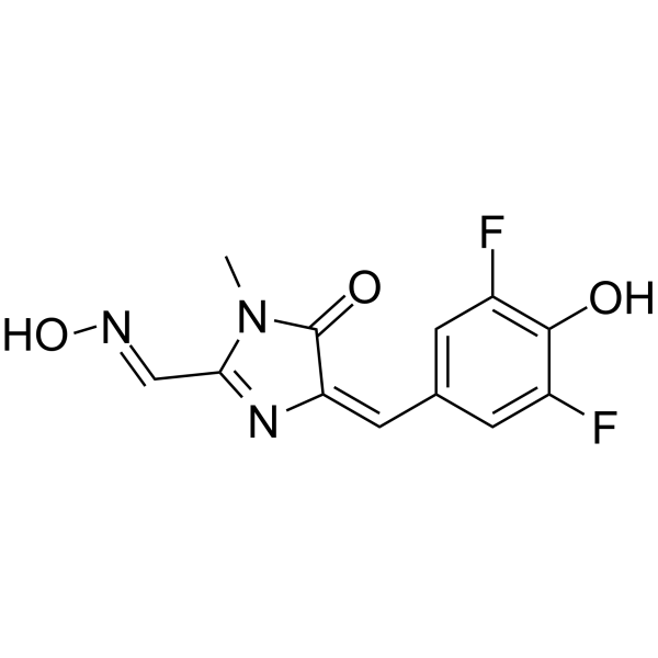
DFHO

DFHO;

DFHO 是 Corn 荧光适体的荧光配体。RNA 适体, Corn 结合 DFHO 的 Kd 值为 70 nM,并将其转化为荧光形式,从而使 RNA 在细胞中成像。

DFHOamp;;

DFHO Chemical Structure

CAS No. : 1420815-34-4

规格 是否有货
1 mg 询价

* Please select Quantity before adding items.

生物活性

DFHO is a fluorogenic ligand of Corn fluorogenic aptamer. The RNA aptamer, Corn binds DFHO with a Kd value of 70 nM and converts it to a fluorescent form, enabling RNA imaging in cells[1][2].

IC50 Target

Kd: 70 nM (Corn binds DFHO)[1]

体外研究
(In Vitro)

Corn is tested in E. coli. Minimal fluorescence that is seen in DFHO-treated cells (20 μM) transformed with the empty vector. However robust yellow fluorescence is seen in E. coli expressing Corn[1].
Corn-DFHO exhibits markedly enhanced photostability in vitro. The Corn-DFHO complexes are imaged in mammalian cells. Since the U6 promoter is one of the three major Pol III promoters, Corn from the U6 promoter is expressed and replaced the majority of the U6 sequence with Corn, leaving a minimal 5′ capping element leader sequence. The majority of the cells expressed diffuse yellow fluorescence, most prominently in the cytoplasm)[1].
DFHO exhibits low background fluorescence and cytotoxicity, making it a potentially suitable fluorophore for activation by RNA aptamers and for imaging experiments in living cells[2].

MCE has not independently confirmed the accuracy of these methods. They are for reference only.

分子量

281.21

Formula

C12H9F2N3O3

CAS 号

1420815-34-4

运输条件

Room temperature in continental US; may vary elsewhere.

储存方式

Please store the product under the recommended conditions in the Certificate of Analysis.

参考文献
  • [1]. Wenjiao Song, et al. Imaging RNA polymerase III transcription using a photostable RNA-fluorophore complex. Nat Chem Biol. 2017 Nov;13(11):1187-1194.

    [2]. Hyaeyeong Kim, et al. A Fluorogenic RNA-Based Sensor Activated by Metabolite-Induced RNA Dimerization. Cell Chem Biol. 2019 Dec 19;26(12):1725-1731.e6.

SCICONS | mRNA疫苗生产中双链RNA (dsRNA)残留检测方案

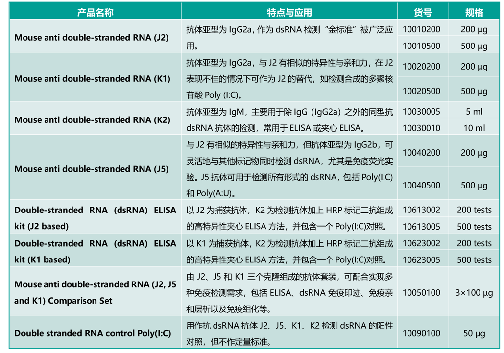
随着mRNA疫苗在抗击COVID-19中的成功应用,针对双链RNA(dsRNA)抗体的需求持续增长。mRNA是DNA通过T7 RNA聚合酶转录生成,dsRNA杂质随之产生,为了监测dsRNA杂质的去除情况,双链RNA (dsRNA) ELISA试剂盒常被应用于mRNA产品的质控环节。

双链RNA (dsRNA) ELISA试剂盒 Double-stranded RNA (dsRNA) ELISA kit

Double-stranded RNA (dsRNA) ELISA kit利用双抗夹心ELISA原理,检测核酸提取物中的病毒dsRNA或非病毒来源的天然或合成dsRNA,以及检测人工合成(m)RNA制剂中的dsRNA杂质。基于使用两种双链RNA(dsRNA)特异性单克隆抗体,Double-stranded RNA (dsRNA) ELISA kit可以灵敏和选择性地检测dsRNA分子(大于40 bp),而不依赖于它们的核苷酸组成和序列,该检测具有高度的特异性。

产品优势:

  • mRNA疫苗中dsRNA杂质残留的监测
  • dsRNA杂质的检测灵敏度高、特异性强
  • 试剂盒适用于检测植物和动物病毒
  • 用于区分细菌病原体和病毒病原体

SCICONS品牌产品

SCICONS是一家向全球研究者提供检测双链RNA(dsRNA)小鼠单克隆抗体的公司,其在1990年就开始利用杂交瘤细胞系生产抗双链RNA(anti-dsRNA)的单克隆抗体,热销的单抗克隆包括J2、K1、K2和J5,这些抗体识别dsRNA的特异性非常强,与DNA或单链RNA ( ssRNA )没有交叉反应,抗体识别具有序列非依赖性,靶向天然存在的长度大于40个碱基对的双链RNA( dsRNA )以及合成的RNA poly ( I :C) 和poly ( A : U )。

SCICONS品牌J2抗体已成为双链RNA(dsRNA)识别的金标准,在国际知名期刊已发表超过200多篇高水平文章。SCICONS于2021年被Nordic-MUbio公司收购,现作为Nordic-MUbio品牌的一个产品线对外销售,产品包括抗体、对照和ELISA检测试剂盒等。

Scicons 双链RNA检测系列产品

货号

英文品名

规格

10010200

Mouse anti double-stranded RNA (J2)

200 µg

10010500

Mouse anti double-stranded RNA (J2)

500 µg

10040200

Mouse anti double-stranded RNA (J5)

200 µg

10040500

Mouse anti double-stranded RNA (J5)

500 µg

10020200

Mouse anti double-stranded RNA (K1)

200 µg

10020500

Mouse anti double-stranded RNA (K1)

500 µg

10050100

Mouse anti double-stranded RNA (J2, J5 and K1) Comparison Kit

3 x 100 µg

10613002

Double-stranded RNA (dsRNA) ELISA kit (J2 based)

Kit

10623002

Double-stranded RNA (dsRNA) ELISA kit (K1 based)

Kit

10623005

Double-stranded RNA (dsRNA) ELISA kit (K1 based)

Kit

10613005

Double-stranded RNA (dsRNA) ELISA kit (J2 based)

Kit

10030010

Mouse anti double-stranded RNA (K2)

10 ml

10030005

Mouse anti double-stranded RNA (K2)

5 ml

SCICONS | mRNA疫苗生产中双链RNA (dsRNA)残留检测方案

J2抗体应用

J2抗体最初用于植物病毒的研究,2006年J2被证明可用于检测所有被正链RNA病毒感染的细胞中产生的dsRNA以来,该抗体在更多领域得到了广泛应用。

  1. J2可用于检测丙型肝炎病毒、登革热病毒、鼻病毒、基孔肯雅病毒、狂犬病病毒、脊髓灰质炎病毒、猪瘟病毒、雀麦花叶病毒等多种病毒的dsRNA中间体。
  2. J2已被用于阐明抗病毒反应是如何启动的,并通过对病毒核酸复制位点进行超微结构定位研究,来探索病毒的生命周期。
  3. J2也被推荐为检测未知病原体是细菌还是病毒的工具。
  4. J2也被用于监测体外合成的m RNA制剂中ds RNA的去除情况。

References

• S. J. Richardson, A. Willcox, D. A. Hilton, S. Tauriain­en, H. Hyoty, A. J. Bone, A. K. Foulis, N. G. Morgan. Use of antisera directed against dsRNA to detect viral infections in formalin-fixed paraffin-embedded tissue. J Clin Virol. (2010) 49(3); 180-5. doi: 10.1016/j. jcv.2010.07.015.

• F. Weber, V. Wagner, S. B. Rasmussen, R. Hartmann, S. R. Paludan. Double-stranded RNA is produced by positive-strand RNA viruses and DNA viruses but not in detectable amounts by negative-strand RNA viruses. J Virol (2006), 80(10):5059-64. doi: 10.1128/ JVI.80.10.5059-5064.2006.

• S. Welsch, S. Miller, I. Romero-Brey, A. Merz, C. K. E. Bleck, P. Walther, S. D. Fuller, C. Antony, J. Kri­jnse-Locker, R. Bartenschlager. Composition and Three-Dimensional Architecture of the Dengue Virus Replication and Assembly Sites. Cell Host & Microbe (2009) 5(4); 365-375. doi.org/10.1016/j. chom.2009.03.007.

• K. Knoops, M. Bárcena, R. W. Limpens, A. J. Koster, A. M. Mommaas, E. J. Snijder. Ultrastructural charac­terization of arterivirus replication structures: re­shaping the endoplasmic reticulum to accommodate viral RNA synthesis. J Virol. (2012) 86(5); 2474-2487. doi:10.1128/JVI.06677-11.

Nordic-MUbio是一家总部设在荷兰的生产和研发高质量免疫学试剂公司,能够为流式细胞术、细胞生物学、免疫学、癌症研究、干细胞等领域提供广泛的产品组合。 2021年4月13日Nordic-MUbio宣布收购Scicons,为世界范围内的研究者提供检测dsRNA的单克隆抗体及相关产品。除此之外,Nordic-MUbio公司供应2000多种用于生物医学和兽医研究的基础免疫学试剂,包括抗血清、抗体、免疫复合物、纯化蛋白、对照品、血清蛋白标准品和其他校准品,包括高度特异性的单克隆抗体,以及用于不同试验体系的标记二抗等。上海金畔是Nordic-MUbio品牌中国一级代理,为客户提供完善的技术支持与售后服务。如对上述产品感兴趣,欢迎随时拨打上海金畔生物科技有限公司客服热线021-50837765或登录上海金畔生物科技有限公司官网www.jinpanbio.cn查询订购。

Self-replicative RNA Reprogramming kit


Self-replicative RNA Reprogramming kit


Stemgent,  ReproCELL集团成员,发布了卓越表现的自我复制RNA重编程试剂盒


YOKOHAMA, 日本–(BUSINESS WIRE)—我们非常高兴的宣布Stemgent Inc. (Cambridge, MA), ReproCELL 集团公司(Yokohama, Japan – JASDAQ: 4978) 发布了新 Stemgent StemRNA™-SR 重编程试剂盒。这是一款可用于血液细胞的重编程的商业化RNA 重编程试剂盒。非病毒,非DNA的重编程试剂盒和自我复制RNA(srRNA)和microRNA技术配套使用,使干细胞研究者更安全,更灵活,更经济,更高效的进行细胞重编程。也可用于人成纤维细胞和血液来源的内皮祖细胞(EPCs)重编程。EPCs基因稳定,可从新鲜和冷冻保存人外周血和脐带血有效建立。该项技术在瑞典斯德哥尔摩举办的的干细胞研究国际学会(ISSCR)第13届年会议时介绍。

“采用非整合RNA重编程技术的Stemgent StemRNA-SR处理病人血液样品,对血液细胞进行重编程是研发临床相关的iPS细胞系重要进步。Joseph Gentile (Chief Executive Officer, Stemgent).


关于 Stemgent ( ReproCELL 集团公司

Stemgent与世界上优秀的干细胞科学家一起研发干细胞研究领域的革新技术和运用。他的使命是简化和支持重编程 Stemgent干细胞试剂提供给全世界的客户。

◆相关阅读


细胞重编程——指南目录


1.    什么是细胞重编程

2.    细胞重编程概述

3.    mRNA重编程

4.    病毒重编程

5.    产品列表 


◆相关产品


货号

名称

规格

00-0073

microRNA Booster Kit

microRNA增强试剂盒

1kit

00-0071

 Stemgent®mRNA Reprogramming Kit

 mRNA 重编程试剂盒

1kit

00-0075

Stemgent® StemRN™ SR Reprogramming Kit

StemRNATM-SR重编程试剂盒

1kit