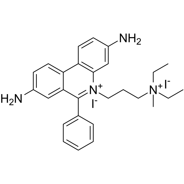
Propidium Iodide;(Synonyms: 碘化丙啶; PI) 纯度: 99.44%
Propidium Iodide 是可用于细胞染色的红色荧光染料。

Propidium Iodide Chemical Structure
CAS No. : 25535-16-4
| 规格 | 价格 | 是否有货 | 数量 |
|---|---|---|---|
| Free Sample (0.1-0.5 mg) | ; | Apply now | ; |
| 10 mg | ¥500 | In-stock | |
| 50 mg | ¥990 | In-stock | |
| 100 mg | ¥1700 | In-stock | |
| 500 mg | ¥5800 | In-stock | |
| 1 g | ; | 询价 | ; |
| 5 g | ; | 询价 | ; |
* Please select Quantity before adding items.
Propidium Iodide 相关产品
bull;相关化合物库:
- Bioactive Compound Library Plus
| 生物活性 |
Propidium Iodide is a red-fluorescent dye that can be used to stain cells. |
||||||||||||||||
|---|---|---|---|---|---|---|---|---|---|---|---|---|---|---|---|---|---|
| 体外研究 (In Vitro) |
Propidium Iodide is a cell-membrane impermeable dye with characteristic excitation maximum at 535 nm and emission maximum at 617 nm which intercalates with nucleic acids with a stoichiometry of one dye per 4-5 base pairs with little sequence preference. Propidium Iodide has evidenced of having no toxic effects on neurons, being today’s most common marker for membrane integrity and cell viability when applied prior to fixation (pre-fixation Propidium Iodide staining method). The pre-fixation staining has been widely used for quantitative assessments of neuronal cell decline in models of acute neurodegeneration, visualized as intensely labeled PI+-pycnotic nuclei of degenerating neurons [1]. Propidium Iodide cannot cross the membrane of live cells, making it useful to measure the percentage of apoptotic cells by flow-cytometric analysis. The flow cytometric data shows an excellent correlation with the results obtained with both electrophoretic and colorimetric methods. This new rapid, simple and reproducible method proves useful for assessing apoptosis of specific cell populations in heterogeneous tissues such as bone marrow, thymus and lymph nodes[2]. MCE has not independently confirmed the accuracy of these methods. They are for reference only. |
||||||||||||||||
| 分子量 |
668.39 |
||||||||||||||||
| Formula |
C27H34I2N4 |
||||||||||||||||
| CAS 号 |
25535-16-4 |
||||||||||||||||
| 中文名称 |
碘化丙啶 |
||||||||||||||||
| 运输条件 |
Room temperature in continental US; may vary elsewhere. |
||||||||||||||||
| 储存方式 |
4deg;C, sealed storage, away from moisture and light *In solvent : -80deg;C, 6 months; -20deg;C, 1 month (sealed storage, away from moisture and light) |
||||||||||||||||
| 溶解性数据 |
In Vitro:;
DMSO : 100 mg/mL (149.61 mM; Need ultrasonic) H2O : 3.57 mg/mL (5.34 mM; ultrasonic and warming and heat to 60°C) 配制储备液
*
请根据产品在不同溶剂中的溶解度选择合适的溶剂配制储备液;一旦配成溶液,请分装保存,避免反复冻融造成的产品失效。 In Vivo:
请根据您的实验动物和给药方式选择适当的溶解方案。以下溶解方案都请先按照 In Vitro 方式配制澄清的储备液,再依次添加助溶剂: ——为保证实验结果的可靠性,澄清的储备液可以根据储存条件,适当保存;体内实验的工作液,建议您现用现配,当天使用; 以下溶剂前显示的百
|
||||||||||||||||
| 参考文献 |
|
| Cell Assay [2] |
Flow cytometric analysis: Propidium iodide is prepared in in 0.1% sodium citrate plus 0.1% Triton X-100 (50 μg/mL). The 200 ×g centrifuged cell pellet is gently resuspended in 1.5 mL hypotonlc fluorochrome solution (Propidium iodide 50 μg/mL), in 12×75 polypropylene tubes. The tubes are placed at 4°C in the dark overnight before the flow cytometric analysis. The propidium Iodide fluorescence of individual nuclei is measured using a FACScan flow cytometer. The nuclei traverses the light beam of a 488 nm Argon laser. A 560 nm dichrolc nurror (DM 570) and a 600 nm band pass filter (bandwidth 35 nm) are used for collecting the red fluorescence due to propidium Iodide staining of DNA and the data are registered on a logarithmic scale[2]. MCE has not independently confirmed the accuracy of these methods. They are for reference only. |
|---|---|
| 参考文献 |
|
