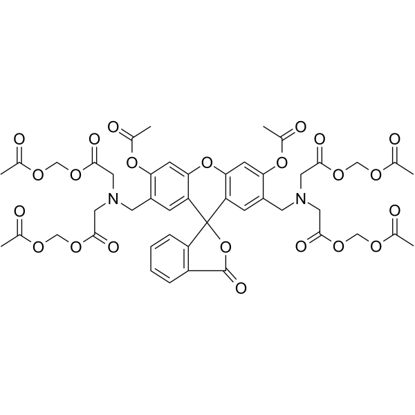
Calcein-AM;(Synonyms: 钙黄绿素-AM; Calcein acetoxymethyl ester) 纯度: ge;96.0%
Calcein-AM是用于测定细胞活力的可以渗透细胞的荧光染料。

Calcein-AM Chemical Structure
CAS No. : 148504-34-1
| 规格 | 价格 | 是否有货 | 数量 |
|---|---|---|---|
| 100 μg(2 mg/mL * 50 μL in DMSO) | ¥1300 | In-stock | |
| 500 μg(2 mg/mL * 250 μL in DMSO) | ¥4750 | In-stock |
* Please select Quantity before adding items.
| 生物活性 |
Calcein-AM is cell-permeable fluorescent dye used to determine the cell viability. |
|---|---|
| 体外研究 (In Vitro) |
The calcein-AM dye used to stain the living cells is shown to have a low spontaneousleakage rate less than 15% in 4 hours at 37°C. Dilutions of targets stained by calcein-AM has a linear relationship with measured fluorescence values. NK cells, LAKs, and CTLs are readily detectable by this microtest. Quantitation of killing and kinetic analysis is readily performed with the test system[1]. Calcein-AM is pH independent, better retained and more photostable. In addition, the high level of intracellular retention of calcein-AM and its low-level release after incorporation exclude possible cell-monolayer labeling and allow its use in a cell-cell interaction assay. Moreover, the bright fluorescence can easily be detected and measured by a microplate fluorescence reader[2]. Calcein-AM is a highly lipophilic vital dye that rapidly enters viable cells, is converted by intracellular esterases to calcein that produces an intense green (530-nm) signal, and is retained by cells with intact plasma membrane. From dying or damaged cells with compromised membrane integrity or from cells expressing multidrug resistance protein (MRP), unhydrolyzed substrates and their fluorescent products are rapidly extruded from cells. The calcein-AM assay has been used to assess the cell viability, cytotoxicity and tp quantitate apoptosis[3]. MCE has not independently confirmed the accuracy of these methods. They are for reference only. |
| 体内研究 (In Vivo) |
Calcein-AM is found to be suitable for in vivo studies, because it has no deleterious effects on cell function and is, indeed, a marker of cell viability[2]. MCE has not independently confirmed the accuracy of these methods. They are for reference only. |
| 分子量 |
994.86 |
| Formula |
C46H46N2O23 |
| CAS 号 |
148504-34-1 |
| 中文名称 |
钙黄绿素-AM |
| 运输条件 |
Room temperature in continental US; may vary elsewhere. |
| 储存方式 |
-20deg;C, protect from light *In solvent : -80deg;C, 6 months; -20deg;C, 1 month (protect from light) |
| 参考文献 |
|
| Cell Assay [1][2][3] |
K562, Daudi, and Chang liver cells are labeled with calcein-AM. Calcein-AM’s excitation and emission wavelengths are 496 nm and 520 nm, respectively. The filter/mirror combination used to detect calcein-AM’s green fluorescence includes the 490-nm excitation and 520-nm emission filters with a dichroic mirror. Differences in the automatic fluorescence readings between the test and control wells determine the results[1]. A simple and sensitive cell-cell adhesion microplate assay is established using the calcein-AM. The procedure involves three steps: the labeling of lymphocytes with an adequate concentration of calcein-AM (20 μM) during a short incubation period (30 min); the adhesion of 2×10⁵ labeled lymphocytes per well to confluent keratinocyte or fibroblast monolayers grown in microtiter plates for 90 min; and, finally, measurement of the fluorescent signal utilizing a new system of cold-light microfluorimetry[2]. Cells are incubated for 15 min in 1 mL of a 1% saponin solution in PBS buffer, pH 7.4, containing 0.05% sodium azide. After saponin permeabilization, 4×10⁵ RBCs in suspension in PBS buffer containing 0.1% saponin and 0.05% sodium azide are incubated (37°C in the dark for 45 min) with calcein-AM to a final concentration of 5 μM, ished three times with the same PBS buffer containing 0.1% saponin and 0.05% sodium azide, and the cell viability is analyzed by flow cytometry[3]. MCE has not independently confirmed the accuracy of these methods. They are for reference only. |
|---|---|
| 参考文献 |
|
