D-Luciferin potassium salt D-荧光素钾盐 品牌:Chemodex


D-Luciferin potassium salt

D-荧光素钾盐

品牌:Chemodex
CAS No.:115144-35-9
储存条件:-20°C
纯度:>95% (HPLC)
产品编号

(生产商编号)

等级 规格 运输包装 零售价(RMB) 库存情况 参考值

CDX-L0009-M050

50 mg 550.00


* 干冰运输、大包装及大批量的产品需酌情添加运输费用


* 零售价、促销产品折扣、运输费用、库存情况、产品及包装规格可能因各种原因有所变动,恕不另行通知,确切详情请联系上海金畔生物科技有限公司。

D-Luciferin potassium salt D-荧光素钾盐 品牌:Chemodex


D-Luciferin potassium salt

D-荧光素钾盐

品牌:Chemodex
CAS No.:115144-35-9
储存条件:-20°C
纯度:>95% (HPLC)
产品编号

(生产商编号)

等级 规格 运输包装 零售价(RMB) 库存情况 参考值

CDX-L0009-M250

250 mg 1,680.00


* 干冰运输、大包装及大批量的产品需酌情添加运输费用


* 零售价、促销产品折扣、运输费用、库存情况、产品及包装规格可能因各种原因有所变动,恕不另行通知,确切详情请联系上海金畔生物科技有限公司。

6-Amino-D-luciferin 6-氨基-D-荧光素 品牌:Chemodex


6-Amino-D-luciferin

6-氨基-D-荧光素

品牌:Chemodex
CAS No.:161055-47-6
储存条件:-20°C
纯度:>99% (HPLC)
产品编号

(生产商编号)

等级 规格 运输包装 零售价(RMB) 库存情况 参考值

CDX-A0306-M001

1 mg 1,060.00


* 干冰运输、大包装及大批量的产品需酌情添加运输费用


* 零售价、促销产品折扣、运输费用、库存情况、产品及包装规格可能因各种原因有所变动,恕不另行通知,确切详情请联系上海金畔生物科技有限公司。

6-Amino-D-luciferin 6-氨基-D-荧光素 品牌:Chemodex


6-Amino-D-luciferin

6-氨基-D-荧光素

品牌:Chemodex
CAS No.:161055-47-6
储存条件:-20°C
纯度:>99% (HPLC)
产品编号

(生产商编号)

等级 规格 运输包装 零售价(RMB) 库存情况 参考值

CDX-A0306-M005

5 mg 2,130.00


* 干冰运输、大包装及大批量的产品需酌情添加运输费用


* 零售价、促销产品折扣、运输费用、库存情况、产品及包装规格可能因各种原因有所变动,恕不另行通知,确切详情请联系上海金畔生物科技有限公司。

D-Luciferin, Sodium Salt(D-萤光素钠盐) 货号: D1007S/D1007L 规格: 10 mg/500 mg

上海金畔生物科技有限公司代理UELANDY荧光染料全系系列产品

D-Luciferin, Sodium Salt(D-萤光素钠盐)

产品货号: D1007S/D1007L

产品规格: 10 mg/500 mg

目录价(元):600/2800

推荐仪器:化学发光仪、小动物活体成像仪、酶标仪

大包装询价


产品概述:

产品参数
外观:可溶于水的浅黄色固体
Ex/Em:328/533 nm
CAS号:103404-75-7
分子式:C11H7N2NaO3S2
分子量:302.3

储存条件
-20℃干燥避光保存,有效期见外包装。

产品介绍 
活体成像技术(optical in vivo imaging)目前主要采用生物发光(bioluminescence)与荧光(fluorescence)两种技术,生物发光法是基于萤光素酶能催化底物化学发光的原理,将体外能稳定表达萤光素酶的细胞株植入动物体内,与后期注射入体内的底物发生反应,利用光学系统检测光强度,间接反映出细胞数量的变化或细胞的定位。这项技术已被广泛应用于多个领域,最常用的有肿瘤或疾病动物模型的建立,并可用于病毒学研究、siRNA研究、干细胞研究、蛋白质相互作用研究等。
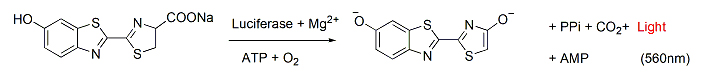
D-Luciferin萤光素酶(Luciferase)的常用底物,普遍用于整个生物技术领域,特别是体内活体成像技术。在ATP萤光素酶的作用下,萤光素能够被氧化,并且在560 nm检测到其化学发光。Luciferinluc基因编码,该基因作为报告基因在多种细胞中存在。由于化学发光的低背景性,luc基因在很低的表达水平下就可以被监测到。此外,萤光素/萤光素酶被用来测量10-15摩尔量的ATP

D-Luciferin, Sodium Salt(D-萤光素钠盐) 货号:               D1007S/D1007L  规格:               10 mg/500 mg


注意事项

1. D-Luciferin , Sodium Salt样品的背景荧光主要来源于萤光素,如果不立即使用本产品,建议分装后,-20℃避光保存。


说明书:

D-Luciferin, Sodium Salt(D-萤光素钠盐) 货号:               D1007S/D1007L  规格:               10 mg/500 mg UE-D1007S/D1007L    
MSDS:

MSDS D1007 D-Luciferin, sodium salt
常见问题解答:

底物的激发波长和发射长?

化学发光,无需激发波长。萤火虫光素发射波长是 560 nm。

推荐仪器?多功能酶标仪能用吗?
1、推荐仪器:具有生物化学发光检测模块。萤光素产生的光可以被光度计或闪烁计数器检测。常见的活体成像仪器:如 IVIS® Lumina 小动物活体成像系统,德国 Bruker公司的 In-Vivo Xtreme 多模式小动物活体成像仪。
2、多功能酶标仪:需要和仪器厂家确认是否具体生物化学发光检测模块。注:不能用荧光显微镜。

底物可以进入活细胞中或者血脑屏障吗?
D-光素约280道尔顿,光素的水溶性和脂溶性都非常好,可以通过血脑屏障,胎盘屏障和血液测试屏障,也可以进入活细胞。

活体成像实验,无效果的原因?
成功进行活体成像实验需要以下条件:
1)目的组织或细胞表达光素酶基因;
2)光素底物注射成功;
3)依赖于发光部位的组织厚度。实验不成功,可从上述因素查找,光素酶基因是否表达,光素底物是否未正确注射,及发光部位较深等

产品不小心放在室温下或暴露在光线下了,还可以用吗?
产品在室温下可以稳定保存很多天,因此产品很可能仍然可以正常工作。 为安全起见,我们建议在处理大量样品或珍贵样品之前,先进行小规模阳性对照实验,以确认该产品仍然适用于您的应用。

两者的区别是什么?
光素钾盐和光素钠盐应用上没有差别,两者的差别在于物理性状上如外形和溶解性。钠盐的水溶性高于钾盐。从目前发表的文献来看,钾盐的使用率远高于钠盐,尤其是体内实验,两者功效相同。

活体成像有必要建立光素酶动力学曲线吗?
注射方式,动物类型以及体重等都会影响信号的发射,因此建议每次实验都要做光素酶动力学曲线,确定最佳信号平台期和最佳的检测时间。


使用本产品的文献:

1.Creating Red Light-Switchable Protein Dimerization Systems as Genetically Encoded Actuators with High Specificity
Zhimin Huang, Zengpeng Li, Xiao Zhang, Shoukai Kang, Runze Dong, Li Sun, Xiaonan Fu, David Vaisar, Kurumi Watanabe,Liangcai Gu
ACS Synth. Biol

参考文献:

1.Application of the firefly luciferase reporter gene
应用方向:体外化学发光分析

2.Optical imaging of Renilla luciferase reporter gene expression in living mice
应用方向:活体成像

3.Real-time measurements of acetylcholine-induced release of ATP from bovine medullary chromaffin cells
应用方向:高灵敏度 ATP 分析

Luciferase-Luciferin, Lyophilized 荧光素-荧光素酶,冻干 品牌:FUJIFILM Wako


Luciferase-Luciferin, Lyophilized

荧光素-荧光素酶,冻干

品牌:FUJIFILM Wako
CAS No.:N/A
储存条件:-20℃
纯度:
产品编号

(生产商编号)

等级 规格 运输包装 零售价(RMB) 库存情况 参考值

123-03921

100 mg

Luciferase-Luciferin, Lyophilized                                                      荧光素-荧光素酶,冻干            品牌:FUJIFILM Wako


* 干冰运输、大包装及大批量的产品需酌情添加运输费用


* 零售价、促销产品折扣、运输费用、库存情况、产品及包装规格可能因各种原因有所变动,恕不另行通知,确切详情请联系上海金畔生物科技有限公司。

D-Luciferin potassium(Synonyms: D-(-)-Luciferin potassium Firefly luciferin potassium Beetle Luciferin potassium)

D-Luciferin potassium;(Synonyms: D-(-)-Luciferin potassium; Firefly luciferin potassium; Beetle Luciferin potassium) 纯度: 99.93%

D-Luciferin (D-(-)-Luciferin) potassium 是萤光素酶的底物,可催化生物发光昆虫产生典型的黄绿光。

D-Luciferin potassiumamp;;(Synonyms: D-(-)-Luciferin potassium; Firefly luciferin potassium; Beetle Luciferin potassium)

D-Luciferin potassium Chemical Structure

CAS No. : 115144-35-9

规格 价格 是否有货 数量
10;mM;*;1 mL in Water ¥660 In-stock
10 mg ¥600 In-stock
50 mg ¥1600 In-stock
100 mg ; 询价 ;
200 mg ; 询价 ;

* Please select Quantity before adding items.

D-Luciferin potassium 相关产品

bull;相关化合物库:

  • Bioactive Compound Library Plus

生物活性

D-Luciferin (D-(-)-Luciferin) potassium is the substrate of luciferases that catalyze the production of light in bioluminescent insects[1].

体外研究
(In Vitro)

D-luciferin is the natural substrate of the enzyme luciferase (Luc), that catalyzes the production of the typical yellowgreen light of fireflies.The present review covers the synthesis of D-luciferin and derivatives or analogues that are substrates or inhibitors of the luciferase from the American firefly Photinus pyralis, the enzyme more frequently used in techniques of in vitro and optical imaging[1].
D-Luciferin exhibits a decrease in the measured Km in PC3M-Luc cell lysates with a Km of 34 μM[2].

MCE has not independently confirmed the accuracy of these methods. They are for reference only.

体内研究
(In Vivo)

Bioluminescence imaging (BLI) using the firefly luciferase (Fluc) as a reporter gene and D-luciferin as a substrate is currently the most widely employed technique. The total signal intensity is plotted against the time after D-luciferin injection to generate a time-intensity curve. In addition to the peak signal, the signals at fixed time points (5, 10, 15, and 20 min) after D-luciferin injection are determined as alternatives to the peak signal. The signal in a given time-intensity curve is normalized for the peak signal in the curve to represent the pattern of temporal changes after D-luciferin injection[3].
Inject with 10 μL of D-luciferin (intraperitoneally or intravenously) stock solution per gram of body weight: normally ~200 μL for a 20 g mouse for a standard 150 mg/kg injection.
Thaw D-Luciferin (either Potassium or Sodium Salt) at room temperature and dissolve in dPBS (no calcium or magnesium) to a final concentration of 15 mg/mL. Pre-wet a 0.22 μm filter by drawing through 5-10 mL of sterile H2O and discard water. Sterilize the D-Luciferin solution through the prepared 0.22 μm syringe filter.

MCE has not independently confirmed the accuracy of these methods. They are for reference only.

分子量

318.41

Formula

C11H7KN2O3S2

CAS 号

115144-35-9

中文名称

D-萤光素钾盐

运输条件

Room temperature in continental US; may vary elsewhere.

储存方式

4deg;C, sealed storage, away from moisture and light

*In solvent : -80deg;C, 6 months; -20deg;C, 1 month (sealed storage, away from moisture and light)

溶解性数据
In Vitro:;

H2O : 25 mg/mL (78.52 mM; Need ultrasonic)

配制储备液
浓度 溶剂体积 质量 1 mg 5 mg 10 mg
1 mM 3.1406 mL 15.7030 mL 31.4060 mL
5 mM 0.6281 mL 3.1406 mL 6.2812 mL
10 mM 0.3141 mL 1.5703 mL 3.1406 mL

*

请根据产品在不同溶剂中的溶解度选择合适的溶剂配制储备液;一旦配成溶液,请分装保存,避免反复冻融造成的产品失效
储备液的保存方式和期限:-80°C, 6 months; -20°C, 1 month (sealed storage, away from moisture and light)。-80°C 储存时,请在 6 个月内使用,-20°C 储存时,请在 1 个月内使用。

参考文献
  • [1]. Giuseppe Meroni, et al. D-Luciferin, derivatives and analogues: synthesis and in vitro/in vivo luciferase-catalyzed bioluminescent activity. ARKIVOC 2009 (i) 265-288.

    [2]. Rajesh Shinde, et al. Luciferin derivatives for enhanced in vitro and in vivo bioluminescence assays. Biochemistry. 2006 Sep 19;45(37):11103-12.

    [3]. Inoue Y, et al. Timing of imaging after d-luciferin injection affects the longitudinal assessment of tumor growthusing in vivo bioluminescence imaging. Int J Biomed Imaging. 2010;2010:471408.

Animal Administration
[2]

Mice[2]
In vivo BLI is performed using a cooled charge-coupled device camera system (IVIS Imaging System 100) 3, 5, 7, 10, 12, 14, 19, 21, 24, and 28 days after the inoculation of HCT116-Luc cells. Mice are injected with 75 mg/kg D-luciferin in 100 μL of phosphate-buffered saline subcutaneously. Beginning 5 min after injection, dorsal luminescent images with an exposure time of 1 s are acquired sequentially at a rate of one image per min until 20 min after D-luciferin injection. Data acquisition is continued until 40 min postinjection on days 3 or 5 and until 25 min on day 7, because of the prolonged time course of light emission. Binning is 4 and the field of view is 15 cm.

MCE has not independently confirmed the accuracy of these methods. They are for reference only.

参考文献
  • [1]. Giuseppe Meroni, et al. D-Luciferin, derivatives and analogues: synthesis and in vitro/in vivo luciferase-catalyzed bioluminescent activity. ARKIVOC 2009 (i) 265-288.

    [2]. Rajesh Shinde, et al. Luciferin derivatives for enhanced in vitro and in vivo bioluminescence assays. Biochemistry. 2006 Sep 19;45(37):11103-12.

    [3]. Inoue Y, et al. Timing of imaging after d-luciferin injection affects the longitudinal assessment of tumor growthusing in vivo bioluminescence imaging. Int J Biomed Imaging. 2010;2010:471408.

D-Luciferin(Synonyms: D-(-)-Luciferin Firefly luciferin Beetle Luciferin)

D-Luciferin;(Synonyms: D-(-)-Luciferin; Firefly luciferin; Beetle Luciferin) 纯度: 99.87%

D-Luciferin (D-(-)-Luciferin) 是萤光素酶的底物,可催化生物发光昆虫产生典型的黄绿光。

D-Luciferinamp;;(Synonyms: D-(-)-Luciferin;  Firefly luciferin;  Beetle Luciferin)

D-Luciferin Chemical Structure

CAS No. : 2591-17-5

规格 价格 是否有货 数量
10 mg ¥600 In-stock
50 mg ¥1600 In-stock
100 mg ; 询价 ;
200 mg ; 询价 ;

* Please select Quantity before adding items.

D-Luciferin 相关产品

bull;相关化合物库:

  • Bioactive Compound Library Plus

生物活性

D-Luciferin (D-(-)-Luciferin) is the substrate of luciferases that catalyze the production of light in bioluminescent insects[1].

体外研究
(In Vitro)

D-luciferin is the natural substrate of the enzyme luciferase (Luc), that catalyzes the production of the typical yellowgreen light of fireflies.The present review covers the synthesis of D-luciferin and derivatives or analogues that are substrates or inhibitors of the luciferase from the American firefly Photinus pyralis, the enzyme more frequently used in techniques of in vitro and optical imaging[1].
D-Luciferin exhibits a decrease in the measured Km in PC3M-Luc cell lysates with a Km of 34 μM[2].

MCE has not independently confirmed the accuracy of these methods. They are for reference only.

体内研究
(In Vivo)

Bioluminescence imaging (BLI) using the firefly luciferase (Fluc) as a reporter gene and D-luciferin as a substrate is currently the most widely employed technique. The total signal intensity is plotted against the time after D-luciferin injection to generate a time-intensity curve. In addition to the peak signal, the signals at fixed time points (5, 10, 15, and 20 min) after D-luciferin injection are determined as alternatives to the peak signal. The signal in a given time-intensity curve is normalized for the peak signal in the curve to represent the pattern of temporal changes after D-luciferin injection[3].
Inject with 10 μL of D-luciferin (intraperitoneally or intravenously) stock solution per gram of body weight: normally ~200 μL for a 20 g mouse for a standard 150 mg/kg injection.
Thaw D-Luciferin (either Potassium or Sodium Salt) at room temperature and dissolve in dPBS (no calcium or magnesium) to a final concentration of 15 mg/mL. Pre-wet a 0.22 μm filter by drawing through 5-10 mL of sterile H2O and discard water. Sterilize the D-Luciferin solution through the prepared 0.22 μm syringe filter.

MCE has not independently confirmed the accuracy of these methods. They are for reference only.

分子量

280.32

Formula

C11H8N2O3S2

CAS 号

2591-17-5

中文名称

D-萤光素

运输条件

Room temperature in continental US; may vary elsewhere.

储存方式

4deg;C, protect from light

*In solvent : -80deg;C, 6 months; -20deg;C, 1 month (protect from light)

溶解性数据
In Vitro:;

DMSO : 50 mg/mL (178.37 mM; Need ultrasonic)

H2O : < 0.1 mg/mL (insoluble)

配制储备液
浓度 溶剂体积 质量 1 mg 5 mg 10 mg
1 mM 3.5674 mL 17.8368 mL 35.6735 mL
5 mM 0.7135 mL 3.5674 mL 7.1347 mL
10 mM 0.3567 mL 1.7837 mL 3.5674 mL

*

请根据产品在不同溶剂中的溶解度选择合适的溶剂配制储备液;一旦配成溶液,请分装保存,避免反复冻融造成的产品失效
储备液的保存方式和期限:-80°C, 6 months; -20°C, 1 month (protect from light)。-80°C 储存时,请在 6 个月内使用,-20°C 储存时,请在 1 个月内使用。

In Vivo:

请根据您的实验动物和给药方式选择适当的溶解方案。以下溶解方案都请先按照 In Vitro 方式配制澄清的储备液,再依次添加助溶剂:

——为保证实验结果的可靠性,澄清的储备液可以根据储存条件,适当保存;体内实验的工作液,建议您现用现配,当天使用; 以下溶剂前显示的百
分比是指该溶剂在您配制终溶液中的体积占比;如在配制过程中出现沉淀、析出现象,可以通过加热和/或超声的方式助溶

  • 1.

    请依序添加每种溶剂:;10% DMSO ;; 90% corn oil

    Solubility: ≥ 2.5 mg/mL (8.92 mM); Clear solution

    此方案可获得 ≥ 2.5 mg/mL (8.92 mM,饱和度未知) 的澄清溶液,此方案不适用于实验周期在半个月以上的实验。

    以 1 mL 工作液为例,取 100 μL 25.0 mg/mL 的澄清 DMSO 储备液加到 900 μL玉米油中,混合均匀。

  • 2.

    请依序添加每种溶剂:;10% DMSO ;; 40% PEG300 ;; 5% Tween-80 ;; 45% saline

    Solubility: ≥ 2.08 mg/mL (7.42 mM); Clear solution

    此方案可获得 ≥ 2.08 mg/mL (7.42 mM,饱和度未知) 的澄清溶液。

    以 1 mL 工作液为例,取 100 μL 20.8 mg/mL 的澄清 DMSO 储备液加到 400 μL PEG300 中,混合均匀;向上述体系中加入50 μL Tween-80,混合均匀;然后继续加入 450 μL生理盐水定容至 1 mL。

    将 0.9 g 氯化钠,完全溶解于 100 mL ddH₂O 中,得到澄清透明的生理盐水溶液

  • 3.

    请依序添加每种溶剂:;10% DMSO ;; 90% (20% SBE-β-CD in saline)

    Solubility: ≥ 2.08 mg/mL (7.42 mM); Clear solution

    此方案可获得 ≥ 2.08 mg/mL (7.42 mM,饱和度未知) 的澄清溶液。

    以 1 mL 工作液为例,取 100 μL 20.8 mg/mL 的澄清 DMSO 储备液加到 900 μL 20% 的 SBE-β-CD 生理盐水水溶液中,混合均匀。

    将 2 g 磺丁基醚 β-环糊精加入 5 mL 生理盐水中,再用生理盐水定容至 10 mL,完全溶解,澄清透明
*以上所有助溶剂都可在 MCE 网站选购。
参考文献
  • [1]. Giuseppe Meroni, et al. D-Luciferin, derivatives and analogues: synthesis and in vitro/in vivo luciferase-catalyzed bioluminescent activity. ARKIVOC 2009 (i) 265-288.

    [2]. Rajesh Shinde, et al. Luciferin derivatives for enhanced in vitro and in vivo bioluminescence assays. Biochemistry. 2006 Sep 19;45(37):11103-12.

    [3]. Inoue Y, et al. Timing of imaging after d-luciferin injection affects the longitudinal assessment of tumor growthusing in vivo bioluminescence imaging. Int J Biomed Imaging. 2010;2010:471408.

Animal Administration
[2]

Mice[2]
In vivo BLI is performed using a cooled charge-coupled device camera system (IVIS Imaging System 100) 3, 5, 7, 10, 12, 14, 19, 21, 24, and 28 days after the inoculation of HCT116-Luc cells. Mice are injected with 75 mg/kg D-luciferin in 100 μL of phosphate-buffered saline subcutaneously near the scapula and were placed in the light-tight chamber of the imaging system. Beginning 5 min after injection, dorsal luminescent images with an exposure time of 1 s are acquired sequentially at a rate of one image per min until 20 min after D-luciferin injection. Data acquisition is continued until 40 min postinjection on days 3 or 5 and until 25 min on day 7, because of the prolonged time course of light emission. Binning is 4 and the field of view is 15 cm.

MCE has not independently confirmed the accuracy of these methods. They are for reference only.

参考文献
  • [1]. Giuseppe Meroni, et al. D-Luciferin, derivatives and analogues: synthesis and in vitro/in vivo luciferase-catalyzed bioluminescent activity. ARKIVOC 2009 (i) 265-288.

    [2]. Rajesh Shinde, et al. Luciferin derivatives for enhanced in vitro and in vivo bioluminescence assays. Biochemistry. 2006 Sep 19;45(37):11103-12.

    [3]. Inoue Y, et al. Timing of imaging after d-luciferin injection affects the longitudinal assessment of tumor growthusing in vivo bioluminescence imaging. Int J Biomed Imaging. 2010;2010:471408.

D-Luciferin sodium(Synonyms: D-萤光素钠盐 D-(-)-Luciferin sodium Firefly luciferin sodium Beetle Luciferin sodium)

D-Luciferin sodium;(Synonyms: D-萤光素钠盐; D-(-)-Luciferin sodium; Firefly luciferin sodium; Beetle Luciferin sodium) 纯度: 99.92%

D-Luciferin (D-(-)-Luciferin) sodium 是萤光素酶的底物,可催化生物发光昆虫产生典型的黄绿光。

D-Luciferin sodiumamp;;(Synonyms: D-萤光素钠盐; D-(-)-Luciferin sodium; Firefly luciferin sodium; Beetle Luciferin sodium)

D-Luciferin sodium Chemical Structure

CAS No. : 103404-75-7

规格 价格 是否有货 数量
10;mM;*;1 mL in DMSO ¥660 In-stock
10 mg ¥600 In-stock
50 mg ¥1600 In-stock
100 mg ; 询价 ;
200 mg ; 询价 ;

* Please select Quantity before adding items.

D-Luciferin sodium 相关产品

bull;相关化合物库:

  • Bioactive Compound Library Plus

生物活性

D-Luciferin (D-(-)-Luciferin) sodium is the substrate of luciferases that catalyze the production of light in bioluminescent insects[1].

体外研究
(In Vitro)

D-luciferin is the natural substrate of the enzyme luciferase (Luc), that catalyzes the production of the typical yellowgreen light of fireflies.The present review covers the synthesis of D-luciferin and derivatives or analogues that are substrates or inhibitors of the luciferase from the American firefly Photinus pyralis, the enzyme more frequently used in techniques of in vitro and optical imaging[1].
D-Luciferin exhibits a decrease in the measured Km in PC3M-Luc cell lysates with a Km of 34 μM[2].

MCE has not independently confirmed the accuracy of these methods. They are for reference only.

体内研究
(In Vivo)

Bioluminescence imaging (BLI) using the firefly luciferase (Fluc) as a reporter gene and D-luciferin as a substrate is currently the most widely employed technique. The total signal intensity is plotted against the time after D-luciferin injection to generate a time-intensity curve. In addition to the peak signal, the signals at fixed time points (5, 10, 15, and 20 min) after D-luciferin injection are determined as alternatives to the peak signal. The signal in a given time-intensity curve is normalized for the peak signal in the curve to represent the pattern of temporal changes after D-luciferin injection[3].
Inject with 10 μL of D-luciferin (intraperitoneally or intravenously) stock solution per gram of body weight: normally ~200 μL for a 20 g mouse for a standard 150 mg/kg injection.
Thaw D-Luciferin (either Potassium or Sodium Salt) at room temperature and dissolve in dPBS (no calcium or magnesium) to a final concentration of 15 mg/mL. Pre-wet a 0.22 μm filter by drawing through 5-10 mL of sterile H2O and discard water. Sterilize the D-Luciferin solution through the prepared 0.22 μm syringe filter.

MCE has not independently confirmed the accuracy of these methods. They are for reference only.

分子量

302.30

Formula

C11H7N2NaO3S2

CAS 号

103404-75-7

中文名称

D-萤光素钠盐

运输条件

Room temperature in continental US; may vary elsewhere.

储存方式

4deg;C, sealed storage, away from moisture and light

*In solvent : -80deg;C, 6 months; -20deg;C, 1 month (sealed storage, away from moisture and light)

溶解性数据
In Vitro:;

H2O : 250 mg/mL (826.99 mM; Need ultrasonic)

DMSO : 100 mg/mL (330.80 mM; Need ultrasonic)

配制储备液
浓度 溶剂体积 质量 1 mg 5 mg 10 mg
1 mM 3.3080 mL 16.5399 mL 33.0797 mL
5 mM 0.6616 mL 3.3080 mL 6.6159 mL
10 mM 0.3308 mL 1.6540 mL 3.3080 mL

*

请根据产品在不同溶剂中的溶解度选择合适的溶剂配制储备液;一旦配成溶液,请分装保存,避免反复冻融造成的产品失效
储备液的保存方式和期限:-80°C, 6 months; -20°C, 1 month (sealed storage, away from moisture and light)。-80°C 储存时,请在 6 个月内使用,-20°C 储存时,请在 1 个月内使用。

In Vivo:

请根据您的实验动物和给药方式选择适当的溶解方案。以下溶解方案都请先按照 In Vitro 方式配制澄清的储备液,再依次添加助溶剂:

——为保证实验结果的可靠性,澄清的储备液可以根据储存条件,适当保存;体内实验的工作液,建议您现用现配,当天使用; 以下溶剂前显示的百
分比是指该溶剂在您配制终溶液中的体积占比;如在配制过程中出现沉淀、析出现象,可以通过加热和/或超声的方式助溶

  • 1.

    请依序添加每种溶剂:;10% DMSO ;; 40% PEG300 ;; 5% Tween-80 ;; 45% saline

    Solubility: ≥ 2.5 mg/mL (8.27 mM); Clear solution

    此方案可获得 ≥ 2.5 mg/mL (8.27 mM,饱和度未知) 的澄清溶液。

    以 1 mL 工作液为例,取 100 μL 25.0 mg/mL 的澄清 DMSO 储备液加到 400 μL PEG300 中,混合均匀;向上述体系中加入50 μL Tween-80,混合均匀;然后继续加入 450 μL生理盐水定容至 1 mL。

    将 0.9 g 氯化钠,完全溶解于 100 mL ddH₂O 中,得到澄清透明的生理盐水溶液

  • 2.

    请依序添加每种溶剂:;10% DMSO ;; 90% (20% SBE-β-CD in saline)

    Solubility: ≥ 2.5 mg/mL (8.27 mM); Clear solution

    此方案可获得 ≥ 2.5 mg/mL (8.27 mM,饱和度未知) 的澄清溶液。

    以 1 mL 工作液为例,取 100 μL 25.0 mg/mL 的澄清 DMSO 储备液加到 900 μL 20% 的 SBE-β-CD 生理盐水水溶液中,混合均匀。

    将 2 g 磺丁基醚 β-环糊精加入 5 mL 生理盐水中,再用生理盐水定容至 10 mL,完全溶解,澄清透明
*以上所有助溶剂都可在 MCE 网站选购。
参考文献
  • [1]. Giuseppe Meroni, et al. D-Luciferin, derivatives and analogues: synthesis and in vitro/in vivo luciferase-catalyzed bioluminescent activity. ARKIVOC 2009 (i) 265-288.

    [2]. Rajesh Shinde, et al. Luciferin derivatives for enhanced in vitro and in vivo bioluminescence assays. Biochemistry. 2006 Sep 19;45(37):11103-12.

    [3]. Inoue Y, et al. Timing of imaging after d-luciferin injection affects the longitudinal assessment of tumor growthusing in vivo bioluminescence imaging. Int J Biomed Imaging. 2010;2010:471408.

Animal Administration
[3]

Mice[3]
In vivo BLI is performed using a cooled charge-coupled device camera system (IVIS Imaging System 100) 3, 5, 7, 10, 12, 14, 19, 21, 24, and 28 days after the inoculation of HCT116-Luc cells. Mice are injected with 75 mg/kg D-luciferin in 100 μL of phosphate-buffered saline subcutaneously near the scapula and were placed in the light-tight chamber of the imaging system. Beginning 5 min after injection, dorsal luminescent images with an exposure time of 1 s are acquired sequentially at a rate of one image per min until 20 min after D-luciferin injection. Data acquisition is continued until 40 min postinjection on days 3 or 5 and until 25 min on day 7, because of the prolonged time course of light emission. Binning is 4 and the field of view is 15 cm.

MCE has not independently confirmed the accuracy of these methods. They are for reference only.

参考文献
  • [1]. Giuseppe Meroni, et al. D-Luciferin, derivatives and analogues: synthesis and in vitro/in vivo luciferase-catalyzed bioluminescent activity. ARKIVOC 2009 (i) 265-288.

    [2]. Rajesh Shinde, et al. Luciferin derivatives for enhanced in vitro and in vivo bioluminescence assays. Biochemistry. 2006 Sep 19;45(37):11103-12.

    [3]. Inoue Y, et al. Timing of imaging after d-luciferin injection affects the longitudinal assessment of tumor growthusing in vivo bioluminescence imaging. Int J Biomed Imaging. 2010;2010:471408.