上海金畔生物科技有限公司代理日本同仁化学 DOJINDO代理商全线产品,欢迎访问官网了解更多信息
特点:
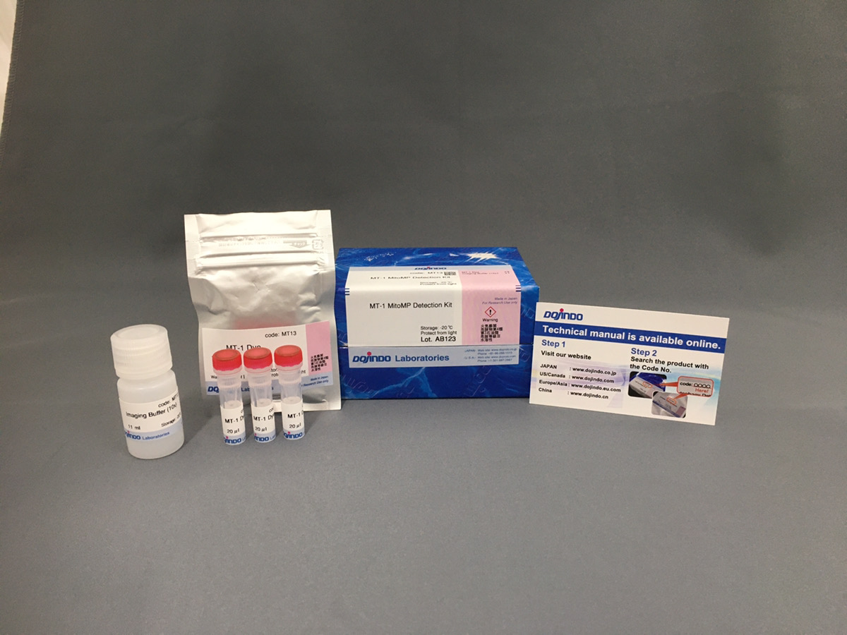
● 只需添加小分子量荧光试剂即可轻松检测线粒体
● 可以使用荧光显微镜进行活细胞成像
● 可以与附着的溶酶体染色剂同时染色
关联产品




上海金畔生物科技有限公司代理日本同仁化学 DOJINDO代理商全线产品,欢迎访问官网了解更多信息
特点:
● 只需添加小分子量荧光试剂即可轻松检测线粒体
● 可以使用荧光显微镜进行活细胞成像
● 可以与附着的溶酶体染色剂同时染色
关联产品


上海金畔生物科技有限公司代理日本同仁化学 DOJINDO代理商全线产品,欢迎访问官网了解更多信息
特点:
● 只需添加小分子量荧光试剂即可轻松检测线粒体
● 可以使用荧光显微镜进行活细胞成像
● 可以与附着的溶酶体染色剂同时染色
注意事项
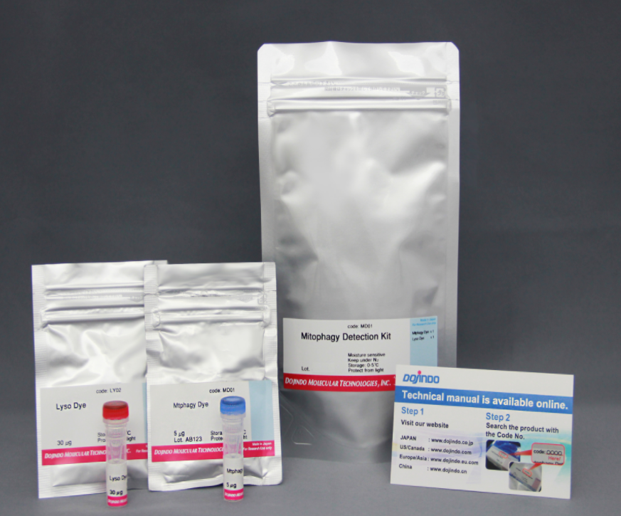
如需检测线粒体自噬,可查看Mitophagy Detection Kit
如您是首次使用Mtphagy染料,建议使用以上试剂盒(内含溶酶体),使用线粒体自噬染料与溶酶体染料共染进行检测
性质
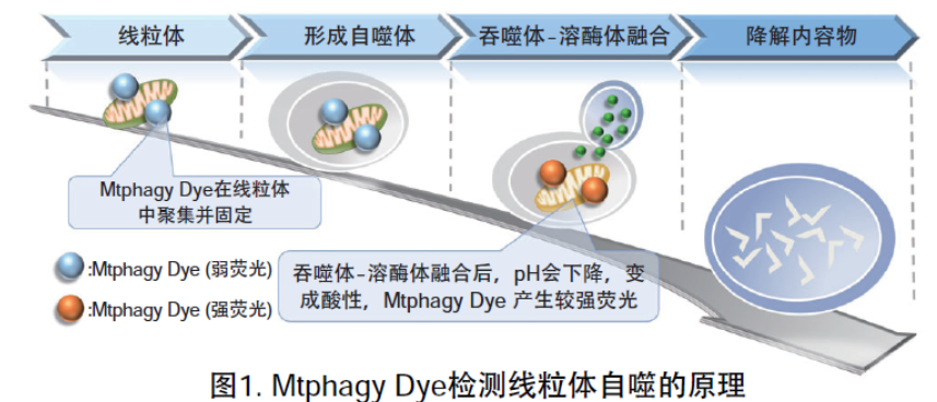
线粒体 (Mitochondria) 是细胞中重要的细胞器之一,可以为细胞活力提供能量 。近年有报道去极化线粒体的积累引起的阿尔茨海默病 (Alzheimer’s Disease) 与帕金森病(Parkinson’s Disease),可能与线粒体自噬有关。线粒体自噬是一种清除机制,可以通过自噬,将氧化应激、DNA损伤因素导致功能失调的线粒体隔离包裹成自噬体(Autophagosome),再与溶酶体 (Lysosome) 融合后降解。本试剂盒内含Mtphagy Dye (用于检测线粒体自噬) 和Lyso Dye (溶酶体染料)。Mtphagy Dye通过化学结合,固定在细胞内的线粒体上,会发出较弱的荧光。当线粒体发生自噬,损伤的线粒体会与溶酶体融合,pH会下降,变成酸性,此时Mtphagy Dye会产生较强的荧光。如想直观观察Mtphagy Dye标记的线粒体和溶酶体的结合,可联合应用试剂盒中的Lyso Dye (标记溶酶体) 进行双染。
首次使用Mtphagy染料时,建议使用包含溶酶体染色试剂(Lyso Dye)的线粒体自噬检测试剂盒(产品货号:MD01),与溶酶体共染色来检测线粒体自噬。
开发者: Dojindo Molecular Technologies, Inc.
检测原理
Mtphagy染料的线粒体自噬检测机制
有关使用Mtphagy染料的实验数据,请参见线粒体自噬—Mitophagy Detection Kit(产品货号MD01)
参考文献
1) J. Koniga, C. Otta, M. Hugoa, T. Junga, A. L. Bulteaub, T. Grunea and A. Hohna, “Mitochondrial contribution to lipofuscin formation”, Redox Biology, 2017, 11, 673.
2) K. Kameyama, “Induction of mitophagy-mediated antitumor activity with folate-appended methyl-β-cyclodextrin”, International Journal of Nanomedicine, 2017, 12, 3433.
3) E. F. Fang, T. B. Waltz, H. Kassahun, Q. Lu, J. S. Kerr, M. Morevati, E. M. Fivenson, B. N. Wollman, K. Marosi, M. A. Wilson, W. B. Iser, D. M. Eckley, Y. Zhang, E. Lehrmann, I. G. Goldberg, M. S. Knudsen, M. P. Mattson, H. Nilsen, V. A. Bohr and K. G. Becker, “Tomatidine enhances lifespan and healthspan in C. elegans through mitophagy induction via the SKN-1/Nrf2 pathway”, Scientific Reports, 2017, 7, (46208), DOI: 10.1038/srep46208.
4) H. Iwashita, S. Torii, N. Nagahora, M. Ishiyama, K. Shioji, K. Sasamoto, S. Shimizu and K. Okuma, “Live Cell Imaging of Mitochondrial Autophagy with a Novel Fluorescent Small Molecule”, ACS Chem. Biol., 2017, 12, (10), 2546.
5) Y. Feng, NB. Madungwe, CV. da Cruz Junho and JC. Bopassa, “Activation of G protein-coupled oestrogen receptor 1 at the onset of reperfusion protects the myocardium against ischemia/reperfusion injury by reducing mitochondrial dysfunction and mitophagy.”, Br. J. Pharmacol., 2017, 174, (23), 4329.
6) K. M. Elamin, K. Motoyama, T. Higashi, Y. Yamashita, A. Tokuda and H. Arima, “Dual targeting system by supramolecular complex of folate-conjugated methyl-β-cyclodextrin with adamantane-grafted hyaluronic acid for the treatment of colorectal cancer.”, Int. J. Biol. Macromol., 2018, doi: 10.1016/j.ijbiomac.2018.02.149.
7)S. Ikeoka and A. Kiso , “The Involvement of Mitophagy in the Prevention of UV-B-Induced Damage in Human Epidermal Keratinocytes “, J. Soc. Cosmet. Chem. Jpn., 2020, 54(3), 252.
规格性状
规格性状:
该产品为紫色至品红色固体。
NMR光谱:测试一致
处理条件
1.保存方法:冷藏,遮光,2.氮气填充,请注意吸潮
关联产品
上海金畔生物科技有限公司代理日本同仁化学 DOJINDO代理商全线产品,欢迎访问官网了解更多信息
特点:
● 只需添加小分子量荧光试剂即可轻松检测线粒体
● 可以使用荧光显微镜进行活细胞成像
● 可以与附着的溶酶体染色剂同时染色
注意事项
如需检测线粒体自噬,可查看Mitophagy Detection Kit
如您是首次使用Mtphagy染料,建议使用以上试剂盒(内含溶酶体),使用线粒体自噬染料与溶酶体染料共染进行检测
性质
线粒体 (Mitochondria) 是细胞中重要的细胞器之一,可以为细胞活力提供能量 。近年有报道去极化线粒体的积累引起的阿尔茨海默病 (Alzheimer’s Disease) 与帕金森病(Parkinson’s Disease),可能与线粒体自噬有关。线粒体自噬是一种清除机制,可以通过自噬,将氧化应激、DNA损伤因素导致功能失调的线粒体隔离包裹成自噬体(Autophagosome),再与溶酶体 (Lysosome) 融合后降解。本试剂盒内含Mtphagy Dye (用于检测线粒体自噬) 和Lyso Dye (溶酶体染料)。Mtphagy Dye通过化学结合,固定在细胞内的线粒体上,会发出较弱的荧光。当线粒体发生自噬,损伤的线粒体会与溶酶体融合,pH会下降,变成酸性,此时Mtphagy Dye会产生较强的荧光。如想直观观察Mtphagy Dye标记的线粒体和溶酶体的结合,可联合应用试剂盒中的Lyso Dye (标记溶酶体) 进行双染。
首次使用Mtphagy染料时,建议使用包含溶酶体染色试剂(Lyso Dye)的线粒体自噬检测试剂盒(产品货号:MD01),与溶酶体共染色来检测线粒体自噬。
开发者: Dojindo Molecular Technologies, Inc.
检测原理
Mtphagy染料的线粒体自噬检测机制

有关使用Mtphagy染料的实验数据,请参见线粒体自噬—Mitophagy Detection Kit(产品货号MD01)
参考文献
1) J. Koniga, C. Otta, M. Hugoa, T. Junga, A. L. Bulteaub, T. Grunea and A. Hohna, “Mitochondrial contribution to lipofuscin formation”, Redox Biology, 2017, 11, 673.
2) K. Kameyama, “Induction of mitophagy-mediated antitumor activity with folate-appended methyl-β-cyclodextrin”, International Journal of Nanomedicine, 2017, 12, 3433.
3) E. F. Fang, T. B. Waltz, H. Kassahun, Q. Lu, J. S. Kerr, M. Morevati, E. M. Fivenson, B. N. Wollman, K. Marosi, M. A. Wilson, W. B. Iser, D. M. Eckley, Y. Zhang, E. Lehrmann, I. G. Goldberg, M. S. Knudsen, M. P. Mattson, H. Nilsen, V. A. Bohr and K. G. Becker, “Tomatidine enhances lifespan and healthspan in C. elegans through mitophagy induction via the SKN-1/Nrf2 pathway”, Scientific Reports, 2017, 7, (46208), DOI: 10.1038/srep46208.
4) H. Iwashita, S. Torii, N. Nagahora, M. Ishiyama, K. Shioji, K. Sasamoto, S. Shimizu and K. Okuma, “Live Cell Imaging of Mitochondrial Autophagy with a Novel Fluorescent Small Molecule”, ACS Chem. Biol., 2017, 12, (10), 2546.
5) Y. Feng, NB. Madungwe, CV. da Cruz Junho and JC. Bopassa, “Activation of G protein-coupled oestrogen receptor 1 at the onset of reperfusion protects the myocardium against ischemia/reperfusion injury by reducing mitochondrial dysfunction and mitophagy.”, Br. J. Pharmacol., 2017, 174, (23), 4329.
6) K. M. Elamin, K. Motoyama, T. Higashi, Y. Yamashita, A. Tokuda and H. Arima, “Dual targeting system by supramolecular complex of folate-conjugated methyl-β-cyclodextrin with adamantane-grafted hyaluronic acid for the treatment of colorectal cancer.”, Int. J. Biol. Macromol., 2018, doi: 10.1016/j.ijbiomac.2018.02.149.
7)S. Ikeoka and A. Kiso , “The Involvement of Mitophagy in the Prevention of UV-B-Induced Damage in Human Epidermal Keratinocytes “, J. Soc. Cosmet. Chem. Jpn., 2020, 54(3), 252.
规格性状
规格性状:
该产品为紫色至品红色固体。
NMR光谱:测试一致
处理条件
1.保存方法:冷藏,遮光,2.氮气填充,请注意吸潮
关联产品


荧光染料Dye Reagents Vat Blue 2;
Vat Blue 2 是一种靛蓝 (HY-N0335) 衍生物,是一种深蓝色的 5,5′-二溴-4,4′-二氯靛染料 (dye)。
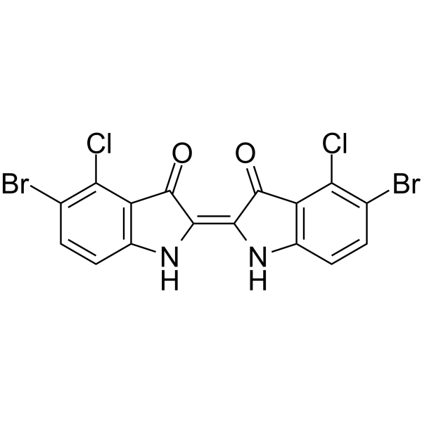
Vat Blue 2 Chemical Structure
CAS No. : 29245-44-1
| 规格 | 是否有货 | ||
|---|---|---|---|
| 5 mg | 询价 | ||
| 10 mg | 询价 | ||
| 100 mg | 询价 |
* Please select Quantity before adding items.
| 生物活性 |
Vat Blue 2, a indigo (HY-N0335) derivative, is a dark blue 5,5′-dibromo-4,4′-dichloroindigo dye[1]. |
|---|---|
| 体外研究 (In Vitro) |
Indigo is the parent compound for a series of bromine and chlorine-substituted dyestuffs. Vat Blue 2 has four substituents, two each of chlorine and bromine, but located at different positions[1]. MCE has not independently confirmed the accuracy of these methods. They are for reference only. |
| 分子量 |
488.95 |
| Formula |
C16H6Br2Cl2N2O2 |
| CAS 号 |
29245-44-1 |
| 运输条件 |
Room temperature in continental US; may vary elsewhere. |
| 储存方式 |
Please store the product under the recommended conditions in the Certificate of Analysis. |
| 参考文献 |
|
荧光染料Dye Reagents Heptamethine cyanine dye-1;(Synonyms: ADS 815EI) 纯度: ge;96.0%
Heptamethine cyanine dye-1是一种用于生物系统荧光成像的近红外花青染料。
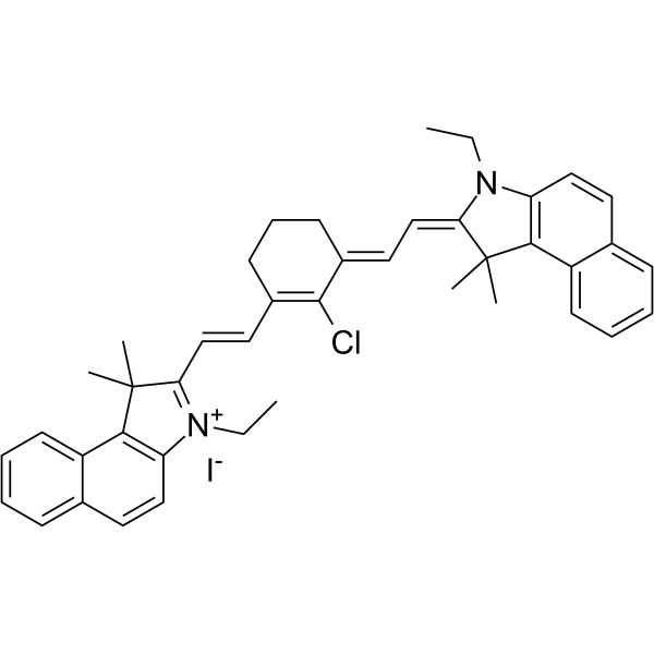
Heptamethine cyanine dye-1 Chemical Structure
CAS No. : 162411-29-2
| 规格 | 价格 | 是否有货 | 数量 |
|---|---|---|---|
| 10;mM;*;1 mL in DMSO | ¥5692 | In-stock | |
| 5 mg | ¥3500 | In-stock | |
| 10 mg | ¥5000 | In-stock | |
| 25 mg | ¥10000 | In-stock | |
| 50 mg | ¥15000 | In-stock | |
| 100 mg | ¥21000 | In-stock | |
| 200 mg | ; | 询价 | ; |
| 500 mg | ; | 询价 | ; |
* Please select Quantity before adding items.
| 生物活性 |
Heptamethine cyanine dye-1 is a near-infrared cyanine dye for fluorescence imaging in biological systems. |
||||||||||||||||
|---|---|---|---|---|---|---|---|---|---|---|---|---|---|---|---|---|---|
| 分子量 |
739.17 |
||||||||||||||||
| Formula |
C42H44ClIN2 |
||||||||||||||||
| CAS 号 |
162411-29-2 |
||||||||||||||||
| 运输条件 |
Room temperature in continental US; may vary elsewhere. |
||||||||||||||||
| 储存方式 |
4deg;C, sealed storage, away from moisture and light *In solvent : -80deg;C, 6 months; -20deg;C, 1 month (sealed storage, away from moisture and light) |
||||||||||||||||
| 溶解性数据 |
In Vitro:;
DMSO : 50 mg/mL (67.64 mM; Need ultrasonic) 配制储备液
*
请根据产品在不同溶剂中的溶解度选择合适的溶剂配制储备液;一旦配成溶液,请分装保存,避免反复冻融造成的产品失效。 In Vivo:
请根据您的实验动物和给药方式选择适当的溶解方案。以下溶解方案都请先按照 In Vitro 方式配制澄清的储备液,再依次添加助溶剂: ——为保证实验结果的可靠性,澄清的储备液可以根据储存条件,适当保存;体内实验的工作液,建议您现用现配,当天使用; 以下溶剂前显示的百
|
荧光染料Dye Reagents NIR dye-1;
NIR dye-1 (Compound 1h) 是一种近红外 (NIR) 荧光染料。NIR dye-1 在 NIR 区域具有吸收和发射作用,同时保留了光学可调的羟基。
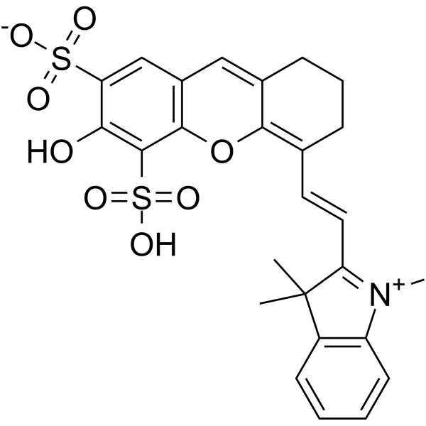
NIR dye-1 Chemical Structure
CAS No. : 1392488-07-1
| 规格 | 是否有货 | ||
|---|---|---|---|
| 100 mg | ; | 询价 | ; |
| 250 mg | ; | 询价 | ; |
| 500 mg | ; | 询价 | ; |
* Please select Quantity before adding items.
| 生物活性 |
NIR dye-1 (Compound 1h) is a near-infrared (NIR) fluorescent dye. NIR dye-1 has absorption and emission in the NIR region, while retaining an optically tunable hydroxyl group[1]. |
|---|---|
| 体外研究 (In Vitro) |
To demonstrate the feasibility of developing water-soluble NIR dyes, NIR dye-1 (Compound 1h) is further synthesized, which bears several water-soluble groups. As expected, dye NIR dye-1 (Compound 1h) is soluble in pure PBS and newborn calf serum (without addition of any co-organic solvent) with a solubility of at least 2 mM in PBS and 3 mM in newborn calf serum[1]. MCE has not independently confirmed the accuracy of these methods. They are for reference only. |
| 分子量 |
543.61 |
| Formula |
C26H25NO8S2 |
| CAS 号 |
1392488-07-1 |
| 运输条件 |
Room temperature in continental US; may vary elsewhere. |
| 储存方式 |
Please store the product under the recommended conditions in the Certificate of Analysis. |
| 参考文献 |
|
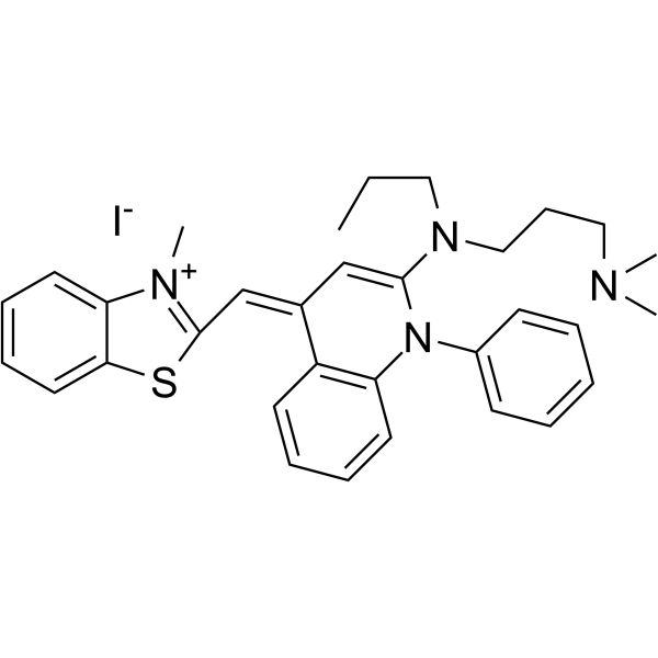
荧光染料Dye Reagents Dye 993;
Dye 993为非对称的花青类染料,有选择渗透性,可用于检测电泳胶中的DNA。

Dye 993 Chemical Structure
| 规格 | 是否有货 | ||
|---|---|---|---|
| 100 mg | ; | 询价 | ; |
| 250 mg | ; | 询价 | ; |
| 500 mg | ; | 询价 | ; |
* Please select Quantity before adding items.
| 生物活性 |
Dye 993, substituted unsymmetrical cyanine dyes with selected permeability, useful in the detection of DNA in electrophoretic gels. |
|---|---|
| 分子量 |
679.70 |
| Formula |
C34H42IN5S |
| 运输条件 |
Room temperature in continental US; may vary elsewhere. |
| 储存方式 |
Please store the product under the recommended conditions in the Certificate of Analysis. |
荧光染料Dye Reagents Quinizarin;(Synonyms: 1,4-二羟基蒽醌; 1,4-Dihydroxyanthraquinone) 纯度: ge;95.0%
Quinizarin (1,4-Dihydroxyanthraquinone) 是 Doxorubicin、Daunorubicin、Adriamycin 等抗癌药物的一部分,通过插入方式与 DNA 相互作用 (Kd=86.1 μM)。Quinizarin 是一种杀菌剂和杀虫剂,并已显示出抑制肿瘤细胞生长的能力。
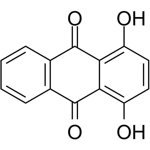
Quinizarin Chemical Structure
CAS No. : 81-64-1
| 规格 | 价格 | 是否有货 | 数量 |
|---|---|---|---|
| 10;mM;*;1 mL in DMSO | ¥550 | In-stock | |
| 1 g | ¥500 | In-stock | |
| 5 g | ; | 询价 | ; |
| 10 g | ; | 询价 | ; |
* Please select Quantity before adding items.
| 生物活性 |
Quinizarin (1,4-Dihydroxyanthraquinone), a part of the anticancer agents such as Doxorubicin, Daunorubicin, and Adriamycin, interacts with DNA by intercalating mode (Kd=86.1 μM). Quinizarin is used as a fungicide and pesticide chemical and has shown the ability to inhibit tumor cell growth[1][2]. |
||||||||||||||||
|---|---|---|---|---|---|---|---|---|---|---|---|---|---|---|---|---|---|
| 体外研究 (In Vitro) |
1,4-Dihydroxyanthraquinone (1,4-DHAQ, a fluorophore) doped cellulose (CL) (denoted as 1,4-DHAQ@CL) microporous nanofiber film has been achieved via simple electrospinning and subsequent deacetylating, and used for highly sensitive and selective fluorescence detection of Cu(2+) in aqueous solution[1]. MCE has not independently confirmed the accuracy of these methods. They are for reference only. |
||||||||||||||||
| 分子量 |
240.21 |
||||||||||||||||
| Formula |
C14H8O4 |
||||||||||||||||
| CAS 号 |
81-64-1 |
||||||||||||||||
| 中文名称 |
1,4-二羟基蒽醌;醌茜 |
||||||||||||||||
| 运输条件 |
Room temperature in continental US; may vary elsewhere. |
||||||||||||||||
| 储存方式 |
|
||||||||||||||||
| 溶解性数据 |
In Vitro:;
DMSO : 3.33 mg/mL (13.86 mM; ultrasonic and warming and heat to 60°C) 配制储备液
*
请根据产品在不同溶剂中的溶解度选择合适的溶剂配制储备液;一旦配成溶液,请分装保存,避免反复冻融造成的产品失效。 |
||||||||||||||||
| 参考文献 |
|
荧光染料Dye Reagents Dye 937;
Dye 937为非对称的花青类染料,有选择渗透性,可用于检测电泳胶中的DNA。
Dye 937 Chemical Structure
CAS No. : 195199-04-3
| 规格 | 是否有货 | ||
|---|---|---|---|
| 100 mg | ; | 询价 | ; |
| 250 mg | ; | 询价 | ; |
| 500 mg | ; | 询价 | ; |
* Please select Quantity before adding items.
| 生物活性 |
Dye 937, substituted unsymmetrical cyanine dyes with selected permeability, useful in the detection of DNA in electrophoretic gels. |
|---|---|
| 分子量 |
636.63 |
| Formula |
C32H37IN4S |
| CAS 号 |
195199-04-3 |
| 运输条件 |
Room temperature in continental US; may vary elsewhere. |
| 储存方式 |
Please store the product under the recommended conditions in the Certificate of Analysis. |
Endoplasmic reticulum dye 1;
Endoplasmic reticulum dye 1 是一种活细胞显像剂 (live cell imaging agent),用于检测质膜上的胞外活动。Endoplasmic reticulum dye 1 具有低细胞毒性,耐光漂白,是短期或长期成像的理想选择。
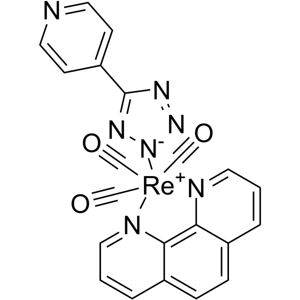
Endoplasmic reticulum dye 1 Chemical Structure
CAS No. : 1404104-40-0
| 规格 | 是否有货 | ||
|---|---|---|---|
| 100 mg | ; | 询价 | ; |
| 250 mg | ; | 询价 | ; |
| 500 mg | ; | 询价 | ; |
* Please select Quantity before adding items.
| 生物活性 |
Endoplasmic reticulum dye 1 is a promising live cell imaging agent for the detection of exocytotic events at the plasma membrane. Endoplasmic reticulum dye 1 shows low cytotoxicity, resistance to photobleaching , which is ideal for imaging either short- or long-time courses[1]. |
|---|---|
| 分子量 |
596.57 |
| Formula |
C21H12N7O3Re |
| CAS 号 |
1404104-40-0 |
| 运输条件 |
Room temperature in continental US; may vary elsewhere. |
| 储存方式 |
Please store the product under the recommended conditions in the Certificate of Analysis. |
| 参考文献 |
|