细胞的能量发电厂——线粒体研究相关产品

细胞的能量发电厂——线粒体研究相关产品

  • 产品特性
  • 相关资料
  • Q&A
  • 参考文献

细胞的能量发电厂——线粒体研究相关产品细胞的能量发电厂——线粒体研究相关产品


线粒体是细胞的动力来源,糖和脂肪在线粒体中被氧化,产生能量,以满足细胞多种功能。线粒体也是活性氧产生、离子稳态和细胞凋亡的主要场所。因此,线粒体功能障碍与代谢、年龄相关的疾病密切相关,如神经退行性疾病、糖尿病、缺血性损伤、癌症等。


为此,Enzo可提供多款产品用于研究代谢调节中的线粒体动力学。更多产品信息,欢迎咨询Enzo在中国的一级代理富士胶片和光。


※ 本页面产品仅供研究用,研究以外不可使用。

产品列表
产品编号 产品名称 产品规格 产品等级 备注
ENZ-51018-0025 MITO-ID®  Membrane potential detection kit
MITO-ID®  膜电位检测试剂盒(荧光显微镜和流式细胞仪)
25 tests
ENZ-51018-K100 MITO-ID®  Membrane potential detection kit
MITO-ID®  膜电位检测试剂盒(荧光显微镜和流式细胞仪)
100 tests
ENZ-51019-KP002 MITO-ID®  Membrane potential cytotoxicity kit
MITO-ID®  膜电位(细胞毒性)试剂盒(微孔板)
1 Kit
ENZ-51007-0100 MITO-ID®  Red detection kit (GFP CERTIFIED) for microscopy
MITO-ID®  红色荧光检测试剂盒(荧光显微镜用)
100 tests
ENZ-51007-500 MITO-ID®  Red detection kit (GFP CERTIFIED) for microscopy
MITO-ID®  红色荧光检测试剂盒(荧光显微镜用)
500 tests
ENZ-51022-0100 MITO-ID®  Green detection kit
MITO-ID®  绿色荧光检测试剂盒(GFP细胞适用)(荧光显微镜用)
100 tests
ENZ-51022-K500 MITO-ID®  Green detection kit
MITO-ID®  绿色荧光检测试剂盒(GFP细胞适用)(荧光显微镜用)
500 tests
ENZ-53007-C200 ORGANELLE-ID-RGB® reagent I
细胞器-ID RGB® 试剂 I
200 µL
ALX-850-276-KI01 Mitochondria/Cytosol fractionation kit
线粒体/胞质分离试剂盒
1 Kit
ALX-850-232-KI01 MitoCapture™ Mitochondrial apoptosis detection kit
MitoCapture™ 线粒体细胞凋亡检测试剂盒
25 tests
ALX-850-232-KI02 MitoCapture™ Mitochondrial apoptosis detection kit
MitoCapture™ 线粒体细胞凋亡检测试剂盒
100 tests
ENZ-51011 ROS-ID®  Total ROS detection kit
总活性氧检测用试剂盒(显微镜和流式细胞仪)
1 Kit

线粒体检测分析

线粒体检测分析

  • 产品特性
  • 相关资料
  • Q&A
  • 参考文献

线粒体检测分析

线粒体检测分析

◆线粒体膜电位检测试剂盒

Mito-ID® Membrane Potential Cytotoxicity Kit – ENZ-51019
  

   灵敏度极高的实时定量线粒体膜电位检测试剂盒。


线粒体检测分析

特点


● 比 JC-1 荧光染料灵敏10倍多

● 试剂盒中含有的染料可发出黄绿双重荧光

 无需漂洗/换液过程

● 适用于高通量筛选


工作原理


试剂盒中含有的染料可发出黄绿双重荧光,在膜电位较高时,染料聚集在线粒体内,呈黄色,同时也以单体形式存在于细胞质中呈绿色;而膜电位较低时,染料主要以绿色单体形式存在于细胞质中,在线粒体内没有黄色。

线粒体膜电位(Mitochondrial Membrane Potential,MMP)是判定细胞健康程度、线粒体膜通透性和细胞凋亡的一个重要指标。线粒体膜电位的下降是早期细胞凋亡的一个标志性事件。此款试剂盒中含有的染料可发出黄绿双重荧光,在膜电位较高时,染料聚集在线粒体内,呈黄色,同时也以单体形式存在于细胞质中呈绿色;而膜电位较低时染料主要以绿色单体形式存在于细胞质中,在线粒体内没有黄色。


产品列表


分类

产品编号

Cellestial产品

应用

细胞类型

染料激发/

发射波长

(nm)

FC

MC

MP

L

F

P


线粒体

ENZ-51018-K100

Mito-ID® Membrane Potential Detection Kit
Mito-ID® 线粒体膜电位检测试剂盒


525/525, 590

ENZ-51019-KP002

Mito-ID® Membrane Potential Cytotoxicity Kit
Mito-ID® 线粒体膜电位细胞毒性试剂盒


525/525 , 590


◆线粒体胞外O2检测试剂盒

Mito-ID® Extracellular O2 Sensor Kit – ENZ-51044
  

氧气敏感型磷光探针可用于培养细胞、单独线粒体、微生物代谢、组织和酶等氧气消耗量的检测。


线粒体检测分析


特点


● 氧气的消耗引起探针磷光信号的增强

● 使用标准的酶标仪(96 或 384 孔板)即可检测,不需要昂贵仪器设备

● 同时也可提供用于胞内氧气检测的渗透性探针

● 可与 Mito-ID® 胞外 pH 检测探针一起使用,验证线粒体特定的毒性


Mito-ID® 胞外氧气检测试剂盒(A)和传统的 ATP 检测方法(B)对线粒体功能的比较,分别用线粒体抑制剂(Oligomycin、Rotenone、Antimycin)、解偶联剂(FCCP)或者对照(DMSO)处理。实验结果表明药物诱导处理后的线粒体功能缺陷立即显现(A 图),而用 ATP 方法检测,经过 24 小时后细胞活力水平差异大(B 图)。


线粒体胞外pH检测探针

Mito-ID® Extracellular pH Sensor Probe – ENZ-51048
    

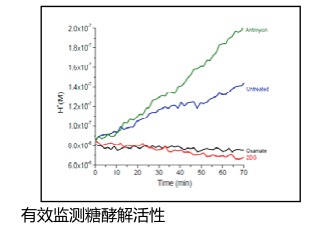
pH 敏感磷光探针可用于监测胞外酸性,而胞外酸性往往是由线粒体毒性致使糖酵解途径增强而产生。

线粒体检测分析


特点


● pH 敏感性探针信号随着酸性的增加而增

● 试剂预混于 96 孔板中,操作简单

● 使用标准的酶标仪即可检测(Ex/Em 340/615nm)


◆HepG2 细胞用葡萄糖转运抑制剂(2DG, Oxamate)或线粒体抑制剂(Antimycin)处理后的酸性变化趋势。


产品编号

产品名称

ENZ-51044

Mito-ID® Extracellular O2 Sensor Kit 
     Mito-ID® 线粒体胞外 O2 检测试剂盒

ENZ-51045

Mito-ID® Extracellular O2 Sensor Kit (High Sensitivity) 
     Mito-ID® 线粒体胞外 O2 检测试剂盒 ( 高灵敏度 )

ENZ-51046

Mito-ID® Intracellular O2 Sensor Probe 
     Mito-ID® 线粒体胞内 O2 检测探针

ENZ-51047

Mito-ID® Extracellular O2 Sensor Probe 
     Mito-ID® 线粒体胞外 O2 检测探针

ENZ-51048

Mito-ID® Extracellular pH Sensor Probe 
     Mito-ID® 线粒体胞外 pH 检测探针


相关产品资料请点击下载:活细胞荧光分析 Ver.3

产品列表
产品编号 产品名称 产品规格 产品等级 备注
多种

高灵敏度的线粒体电位染料


高灵敏度的线粒体电位染料

高灵敏度的线粒体电位染料

  线粒体在细胞代谢,生物能量和细胞凋亡中起着重要的作用。线粒体功能的降低是各种器官中药物相关毒性的主要原因。在早期药物开发过程中,评价药物的细胞毒性作用至关重要。

  Enzo生产的MITO-ID® 线粒体膜电位(细胞毒性)试剂盒,利用阳离子双发射染料检测线粒体(MMP)的波动。


◆特点


● 比JC-1灵敏10倍

● 能在较低药物/剂量浓度检测毒性

● 适合时相研究

● 无清洗和无介质去除

● 兼容高通量应用

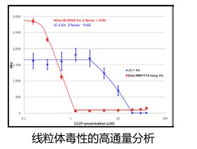
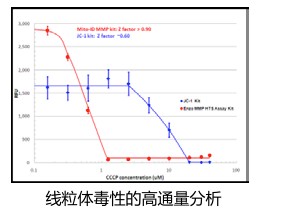
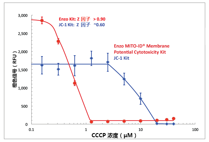
MITO-ID® 表现出出色的灵敏度

高灵敏度的线粒体电位染料

线粒体膜电位降低在CCCP浓度函数中显示为橙色信号的下降。MITO-ID® 膜电位染料

比常用染料JC-1对线粒体电位损失的敏感性至少高10倍。


  Enzo为早期安全性高通量评估提供了全套工具,包括SCREEN-WELL® 毒性库,基于细胞的检测,如凋亡氧化应激细胞活性等。(详细可点击文字)

产品列表

产品编号

产品名称

包装

ENZ-51019-KP002

线粒体膜电位(细胞毒性)试剂盒(微孔板)

Mito-ID® Membrane potential cytotoxicity   kit for microplates

1 Kit

ENZ-51018-K100

线粒体膜电位检测试剂盒(流式和荧光显微镜)

Mito-ID® Membrane potential detection kit   for microscopy and flow cytometry

100 tests

JC-1(Synonyms: CBIC2)

JC-1;(Synonyms: CBIC2) 纯度: ge;99.0%

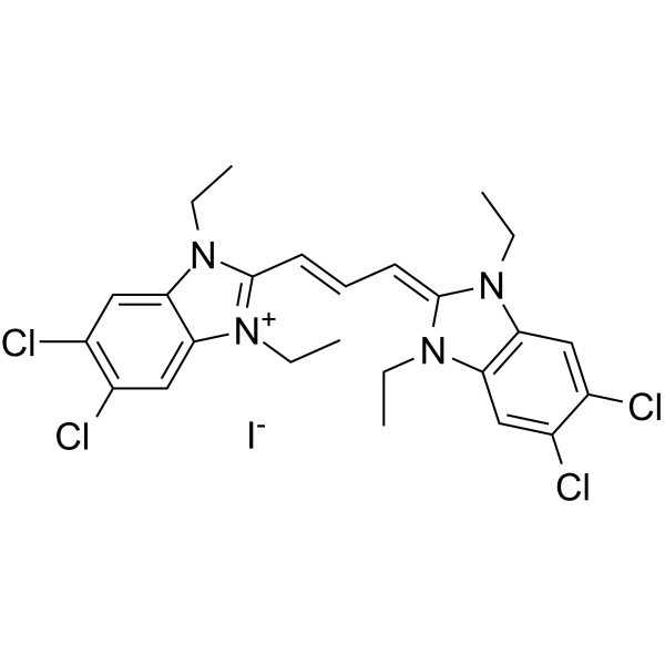
JC-1 (CBIC2) 是荧光亲脂性羰花青染料,用于测量线粒体膜电位。线粒体膜电位较高时, JC-1 在基质中汇聚形成聚合物 (J-aggregates),可以产生红色荧光 (Ex/Em=585/590 nm);线粒体膜电位较低时,JC-1 不能聚集在线粒体基质中,以单体形式存在产生绿色荧光 (Ex/Em=510/527 nm)。

JC-1amp;;(Synonyms: CBIC2)

JC-1 Chemical Structure

CAS No. : 3520-43-2

规格 价格 是否有货 数量
1 mg ¥700 In-stock
2 mg ¥1200 In-stock
5 mg ¥2400 In-stock
10 mg ¥4100 In-stock
50 mg ¥12899 In-stock
100 mg ; 询价 ;
200 mg ; 询价 ;

* Please select Quantity before adding items.

JC-1 相关产品

bull;相关化合物库:

  • Bioactive Compound Library Plus

生物活性

JC-1 (CBIC2) is a fluorescent lipophilic carbocyanine dye used to measure mitochondrial membrane potential. JC-1 forms complexes known as J-aggregates at high ΔΨm. Aggregates of JC-1 emit an orange-red fluorescence (Ex/Em=585/590 nm). While in cells with low ΔΨm, JC-1 remains in the monomeric form. JC-1 monomers emit a green fluorescence (Ex/Em=510/527 nm).

体外研究
(In Vitro)

Guidelines (Following is our recommended protocol. This protocol only provides a guideline, and should be modified according to your specific needs).
Labeling of Cells:
1. Culture cells in 6-, 12- , 24-, or 96-well plates at a density of 5× 10⁵ cells/mL. Incubate the cells according to your normal protocol.
2. Ensure that the JC-1 and DMSO has equilibrated to room temperature, and then prepare a 200 μM stock solution by dissolving the contents of one vial in DMSO provided.
3. For the control tube, allow the vial of CCCP has come to room temperature, add 1 μL of CCCP (50 mM). Incubate cells at 37°C for 5 minutes.
4. Add 10 μL JC-1 (200 μM) per well to make the final concentration at 2 μM. Incubate cells at 37°C, 5% CO2, for 15-20 minutes. If additional labeling followed, for example with an annexin V, begin with step 2.a. If not, proceed with step 1.e.
5. After incubation, centrifuge cells for 3-4 minutes at 400× g at 4°C, carefully aspirate the supernant.
6. Wash cells twice with PBS (1×): add 2 mL PBS (1×) to suspend cells and vortex to mix thoroughly. Centrifuge cells for 3-4 minutes at 400× g at 4°C, carefully aspirate the supernant.
7. Add 500 μL PBS (1×) to suspend cells. Analyze sample on a flow cytometer, fluorescence microscopy, or fluorescence microplate reader.

MCE has not independently confirmed the accuracy of these methods. They are for reference only.

分子量

652.23

Formula

C25H27Cl4IN4

CAS 号

3520-43-2

运输条件

Room temperature in continental US; may vary elsewhere.

储存方式

4deg;C, sealed storage, away from moisture and light

*In solvent : -80deg;C, 6 months; -20deg;C, 1 month (sealed storage, away from moisture and light)

溶解性数据
In Vitro:;

DMSO : 5 mg/mL (7.67 mM; ultrasonic and warming and heat to 60°C)

H2O : < 0.1 mg/mL (insoluble)

配制储备液
浓度 溶剂体积 质量 1 mg 5 mg 10 mg
1 mM 1.5332 mL 7.6660 mL 15.3320 mL
5 mM 0.3066 mL 1.5332 mL 3.0664 mL
10 mM

*

请根据产品在不同溶剂中的溶解度选择合适的溶剂配制储备液;一旦配成溶液,请分装保存,避免反复冻融造成的产品失效
储备液的保存方式和期限:-80°C, 6 months; -20°C, 1 month (sealed storage, away from moisture and light)。-80°C 储存时,请在 6 个月内使用,-20°C 储存时,请在 1 个月内使用。

In Vivo:

请根据您的实验动物和给药方式选择适当的溶解方案。以下溶解方案都请先按照 In Vitro 方式配制澄清的储备液,再依次添加助溶剂:

——为保证实验结果的可靠性,澄清的储备液可以根据储存条件,适当保存;体内实验的工作液,建议您现用现配,当天使用; 以下溶剂前显示的百
分比是指该溶剂在您配制终溶液中的体积占比;如在配制过程中出现沉淀、析出现象,可以通过加热和/或超声的方式助溶

  • 1.

    请依序添加每种溶剂:;10% DMSO ;; 40% PEG300 ;; 5% Tween-80 ;; 45% saline

    Solubility: 1.25 mg/mL (1.92 mM); Suspended solution; Need ultrasonic

    此方案可获得 1.25 mg/mL (1.92 mM) 的均匀悬浊液,悬浊液可用于口服和腹腔注射。

    以 1 mL 工作液为例,取 100 μL 12.5 mg/mL 的澄清 DMSO 储备液加到 400 μL PEG300 中,混合均匀;向上述体系中加入50 μL Tween-80,混合均匀;然后继续加入 450 μL生理盐水定容至 1 mL。

    将 0.9 g 氯化钠,完全溶解于 100 mL ddH₂O 中,得到澄清透明的生理盐水溶液

  • 2.

    请依序添加每种溶剂:;10% DMSO ;; 90% (20% SBE-β-CD in saline)

    Solubility: 1.25 mg/mL (1.92 mM); Suspended solution; Need ultrasonic

    此方案可获得 1.25 mg/mL (1.92 mM) 的均匀悬浊液,悬浊液可用于口服和腹腔注射。

    以 1 mL 工作液为例,取 100 μL 12.5 mg/mL 的澄清 DMSO 储备液加到 900 μL 20% 的 SBE-β-CD 生理盐水水溶液中,混合均匀。

    将 2 g 磺丁基醚 β-环糊精加入 5 mL 生理盐水中,再用生理盐水定容至 10 mL,完全溶解,澄清透明
*以上所有助溶剂都可在 MCE 网站选购。
参考文献
  • [1]. A Perelman, et al. JC-1: alternative excitation wavelengths facilitate mitochondrial membrane potential cytometry. Cell Death Dis. 2012 Nov 22;3:e430.

    [2]. Vera C. Keil, et al. Ratiometric high-resolution imaging of JC-1 fluorescence reveals the subcellular heterogeneity of astrocytic mitochondria. Pflügers Archiv – European Journal of Physiology. 2011,462(5): 693-708.

    [3]. Jung-Ho LEE, In-Hwan LEE, Young-Jun CHOE, et al. Real-time analysis of amyloid fibril formation of α-synuclein using a fibrillation-state-specific fluorescent probe of JC-1. Biochem. J. 2009, 418:311-323.

    [4]. Salvioli S, et al. JC-1, but not DiOC6(3) or rhodamine 123, is a reliable fluorescent probe to assess delta psi changes in intact cells: implications for studies on mitochondrial functionality during apoptosis. FEBS Lett. 1997 Jul 7;411(1):77-82.